Parts of the Microscope
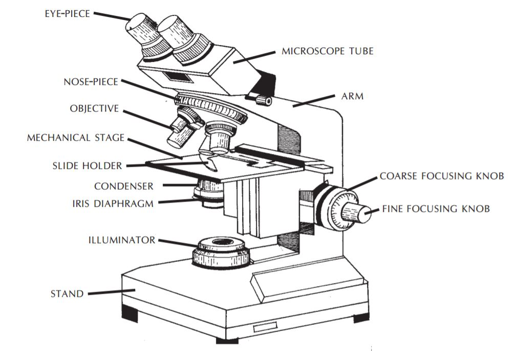
The main parts of the microscope are the eye-pieces, microscope tube, nose-piece, objective, mechanical stage, condenser, coarse and fine focusing knobs, and light source.
`text(Eye-pieces)`
The specimen is viewed through the eye-piece. It has a lens which magnifies the image formed by the objective. The magnifying power of the eye-piece is in the range `text(5x-20x)`. A movable pointer may be attached to the inside of the eye-piece.
In binocular microscopes, the two eye-pieces can be moved closer or farther apart to adjust for the distance between the eyes by pulling pushing motion or by moving a knurled ring.
`text(Microscope tube)`
The microscope tube is attached on top of the arm. It can be of the monocular or binocular type. It supports the eye-piece on the upper end.
`text(Mechanical tube length)`
Mechanical tube length is the distance between the place where the objective is inserted and the top of the draw-tube into which the eyepieces fit.
In modern microscopes it is not tubular; it contains prisms that bend the light coming up, thus providing a comfortable viewing angle. In a binocular tube, the light is also split and sent to both eye-pieces.
`text(Nose-piece)`
The nose-piece is attached under the arm of the microscope tube. The nose-piece houses the objectives and rotates them.
The objectives are arranged in sequential order of their magnifying power, from lower to higher. This helps to prevent the immersion oil from getting onto the intermediate objectives.
`text(Objectives)`
The image of the specimen first passes through the objective.
Objectives with magnifying powers 4x, 10x, 40x and 100x are commonly used.
The magnifying power is marked on the lens and is usually colour-coded for easy identification.
`text(The 100x objective is for oil immersion.)`
The numerical aperture (NA) is the measure of light-gathering power of a lens. The NA corresponding to the various magnifying powers of the objective is:
`text(Magnification Numerical aperture)`
`text( 10x 0.25)`
`text( 40x 0.65)`
`text( 100x 1.25)`
A high NA indicates a high resolving power and thus useful magnification.
To provide the best image at high magnification, immersion oil is placed between the slide and the oil immersion objective (100x). Unlike air, immersion oil has the same refractive index as glass. Therefore, it improves the quality of the image. If immersion oil is not used, the image appears blurred or hazy.
`text(Mechanical stage)`
The mechanical stage holds the slide and allows it to be moved to the left, right, forward or backward by rotating the knobs.
It is fitted with fine vernier graduations as on a ruler.
This helps in relocating a specific field of examination.
`text(Condenser)`
The condenser illuminates the specimen and controls the amount of light and contrast. There are different types of condensers. Some condensers have a rack-and pinion mechanism for up-and-down adjustment.
The NA of a condenser should be equal to or greater than that of the objective with maximum NA.
An iris diaphragm is provided below the condenser. This adjusts the NA of the condenser when using objectives having low magnifying power.
A swing-out type filter holder may be fitted above or under the condenser. In some microscopes the filter holder may not be swing-out type. The filter holder holds detachable filters when required.
Condenser centering screws, when present, are used to align the condenser with the objective.
A condenser raising knob may be present (if centering screws are not there), or the distance may be fixed.
`text(Two-sided mirror)`
A mirror is the simplest illuminator.
The two-sided mirror provides necessary illumination through reflection of natural or artificial light.
It has two surfaces, one plain for artificial light and other concave for natural light.
It is supported on two sides by a fork fixed on a mount in a way that permits free rotation.
`text(A mirror is usually fitted on a mount or at the base of the microscope.)`
`text(Built-in light sources)`
An illuminator is built into the base of the microscope. A halogen bulb provides the best illumination. On top of the illuminator is an in-built filter holder to fit the filter of desired quality.
`text(Filters)`
Blue filters are used to change the light from ordinary electric bulbs into a more natural white light.
Neutral density filters are used to reduce brightness without changing the colour of the background.
Green filters may be useful in some situations.
`text(Immersion oil)`
Immersion oil must be used with objectives having NA more than 1.0. This increases the resolving power of the objective.
An immersion oil of medium viscosity and refractive index of 1.5 is adequate.
`text(Coarse and fine focusing knobs)`
The coarse and fine focusing knobs are used to change the distance between the specimen slide and the objective.
The coarse focusing knob alters this distance rapidly and is used to bring the specimen into the field of view using an objective having low magnification power.
The fine focusing knob changes the distance very slowly and permits better viewing of the object.
One revolution of the fine focusing knob should generally move the mechanical stage by 100 µm. The movement should be smooth and free from jerks.
`text(Halogen lamp)`
Halogen lamps are low wattage, high intensity lamps and are the preferred light source. Though costlier, these have the following advantages over tungsten lamps:
`=>` emit white light
`=>` have higher luminosity (brighter)
`=>` have compact filament
`=>` have longer life.