Biology REVISION OF Structural organisation of plants and animals FOR NDA

PLANT MORPHOLOGY

• Morphology is the branch of biology dealing with the study of form and structure of organisms and their
specific structural features. It includes the study of external structure such as root, stem, leaves, etc.

• On the basis of external appearance, plants can be of following types

(i) Herbs, e.g. carrot, turmeric, pea, etc.

(ii) Shrubs, e.g. rose, sunflower, etc.

(iii) Trees, e.g. neem, mango, bamboo, etc.

Root

In plants, root is the non-green (due to the absence of chlorophyll), cylindrical and descending part that normally
grows downwards into the soil.

It develops from the radicle of embryo. It docs not bear leaves, buds and not distinguished into nodes and
internodes. In some roots, buds are found for vegetative propagation, e.g. sweet potato.

There are two types of roots

(i) Tap Roots Tap root is the primary root that

develops directly through radicle. It bears secondary

and tertiary roots, root caps and root hairs. The primary root with secondary, tertiary roots
constitutes tap root system, e.g. most dicots.

(ii) Adventitious Roots Roots that develop from any part of the plant other than the radicle arc called as
adventitious roots, e.g. grass, Monstera, banyan tree, etc.

Modifications of Tap Root

(i) Napiform These become very thick at the base and tapers towards the apex, e.g. turnip, sugarbeet, etc.

(ii) Fusiform These roots become thicker in middle and tapers at both the ends, e.g. radish (Raphanus
sativus).

(iii) Conical Swollen at base and narrow at apex,
e.g. carrot.

Modifications of Adventitious Roots
For the Stroage of Food

• Tuberous roots From the nodes of the stem, swollen without any defnite shape, e.g. sweet potato.

• Fasciculated roots Arise in bunches, e.g. Asparagus, Dahlia.

• Nodulose roots Apical portion swells up,
e.g. Curcuma, etc.

• Annular roots Ring structure formed, e.g. Psychotria.

For Support

• Prop or pillar roots Hang from branches and penetrate into soil, e.g. banyan, screwpine.
Stilt or brace roots Develop from lower nodes of stem to give additional support, e.g. maize, sugarcane, etc.

• Climbing roots Arise from nodes and help in climbing, e.g. Pothos, Piper betle.

• Buttress roots Arise from basal part of main stem, e.g. Ficus.

• Contractile roots Underground and fleshy, help the plant in fixation, e.g. onion, corm of Crocus, etc.
For Vital Functions

• Floating roots Arise from nodes, help in floating, e.g.] ussiaea.

• Photosynthetic or assimilatory roots Have chlorophyll, e.g. Trapa, Tinospora.

• Reproductive roots Develop vegetative buds,

e.g. Trichosanthes dioica.

• Mycorrhizal roots With fungal hyphae, e.g. Pinus.

•Thorn roots Serves as protective organ, e.g. Pothos.

Functions of Roots

The functions of roots arc given below

(i) Roots anchor the plant from the substratum and perform very important function of absorption of
water and minerals from the soil.

(ii) Roots hold the soil particles firmly to prevent soil erosion.

(iii) Roots also perform some secondary functions with the help of its modification like food storage,
additional mechanical support, act as haustoria, reproduction and nitrogen-fixation.
Stem

It is ascending part of plant and formed by the prolongation of the plumule of embryo. It is positively
phototropic and negatively geotropic and hydrotropic. It bears nodes and internodes. In flowering plants, stem
bears leaves, branches (arise from the cortex), flowers and fruits. Leaf bearing part of stem is called shoot.

Modifications of Stems

Stems usually modify to perform following functions

(i) Survival in the adverse conditions (perennation).

(ii) For helping in vegetative propagation.

(iii) For storage of food.

Modification of Underground Stem

• Rhizome occurs underground, gives rise to annual aerial branches or leaves, e.g. Collocasia, Amcnphophallus,
ginger, turmeric.

• Bulb is underground condensed shoot having a reduced, discoid stem with fleshy scales, e.g. garlic, onion, tulips,
lilies, etc.

• Tuber is swollen tips of underground branches, e.g. potato (Solanum tuberosum ).
Corms are swollen underground vertical stems bearing buds and membranous leaves on upper surface and
adventitious roots below, e.g. Colocasia, Gladiolus, Ammphophallus, Colchicum, etc.

Modifications of Subaerial Stem

• Runners are specia:, narrow, green, horizontal branches, which develop at tbe base of crown, e.g. grasses.

• Stolons arc underground, e.g. Coloc.asia or above ground, e.g. strawb.:rry horizontal branches, which
develop at the base of a crown.

• Offsets are one internode long runners formed in rosette plants at ground or water level, e.g. water lettuce (Pistia).

• Suckers are non-green narrow stem, develops at the

underground base of a crown grows horizontally,
e.g. Mentha (mint), roses, etc.

Modifications of Aerial Stem

• Phylloclades arc gr·:en, photosynthetic, often succulent stems, e.g. Opunl it1

• Cladodes are green stems with leaves reduced to scales or modified into spines and limited growth, e.g. Ruscu.s,
Asparagus.

• Bulbi! A multicellular structure, functions as organ of vegetative reproduction, e.g. Oxalis, Dioscorea, etc.

• Stem thorn Axil o: the leaf or apex of the branch is modified into point eel structure called thorn, e.g. Citrus,
Bougainvillea, etc.

• Stem tendril In plants with weak stem, the apical bud is modified into tendril for climbing, e.g. Passiflora,
cucumber, etc.

Functions of Stem

Functions of stem arc given below

(i) The stem supports the leaves, branches, flowers, fruits and conducts water and minerals from the roots to
the leaves and svnthesiscd food from the leaves to other plant parts.

(ii) It also bears flowers and fruits.

(iii) It performs various secondary functions like storage, vegetative propagation and support.

Leaf

The leaf is green (dut to the presence of photosynthetic pigment chlorophyll ), flat, thin and expanded. It is a
hteral appendage of stem, which is borne at a node and hears a bud in its axil.

On the basis of incision of lamina, leaves are of

two types

(i) Simple leaves In this, there is a single lamina, which is entire, i.e. no incisions are
present, e.g. mango, guava, Cu.curbita, etc.

(ii) Compound leaves In these leaves lamina is divided completely into distinct and separate
segments called leaflets. Rachis is the main axis on which leaflets are arranged.


These are mainly of two types

(a) Palmate compound leaf is one, in which the petiole bears leaflets at the tip, like
the fingers of the palm, e.g. Bombax.

(b) In pinnately compound leaf, leaflets are borne laterally on an elongated axis. It is
of various kinds.

Venation in Leaves

The arrangement of veins and veinlets in leaf lamina is known as venation. It is of following
types

(i) Reticulate venation The veinlets are irregularly distributed to form a network,
e.g. dicot plants. Callophyllum is the dicot plant with parallel venation.


(ii) Parallel venation The veins are arranged parallel to each other, e.g. monocots. Smilax
and Dioscorea are monocots having reticulate venation.

Phyllotaxy

Phyllotaxy is the arrangement of leaves on the stem or its branches. Function of phyllotaxy is to
arrange leaves in such a way that all of them get proper exposure to sunlight.

(i) In alternate type of phyllotaxy a single leaf arises at each node in alternate manner, e.g.
Mangifera indica (mango), Hibiscus rosa sinensis (China rose), Brassica campestris
(mustard), Nicotiana tabacum (tobacco).

(ii) In opposite type of pbyllotaxy, each node gives rise to two leaves lying opposite to
each other, e.g. Calotropis, guava. Opposite phyllotaxy may be either opposite
superimposed (i.e. position of two leaves of each node resembles with the leaves of upper
node), e.g. Eugenia, Quisqualis, lxora, etc., or opposite decussate, (i.e. leaves of a node are
at right angle to the leaves of next node), e.g. Calotropis procera, Ocimum, etc. It should be
noted that in guava (Psidium guavava) both

types of arrangements are found.

(iii) In whorled phyllotaxy, more than two leaves arise at a node and form a whorl, e.g.
Alstonia, Nerium, Vangueria, etc.

Modifications of Leaves

• Leaf tendrils Leaf or leaf parts are modified into thread-like sensitive structure called tendrils, e.g. Lathyrus odoratus.

• Leaf spines Leaf parts are changed into spines in order to protect the plant form grazing animals and excessive transpiration, e.g.
Alee.

• Ph:vllodes Flattened green, photosynthetic, petioles and rachis, e.g Utricularia, Acacia.

• Led pitchers Lamina or whole leaf is modified into pitcher, e.g. Nepenthes.

• Succulent leaves are fleshy or swollen, e.g. Aloe, Agave.

• Stroage leaves are swollen, e.g. Allium (onion).

• Xerophytic leaves usually have hard, waxy tiny leaves that are usually modified into thorns to reduce water loss.

Functions of Leaves

Leaves in plants perform several functions as given below

(i) Leaves carry out photosynthesis and possess stomata for the gaseous exchange and transpiration.

(ii) The leaves protect terminal and axillary buds.

(iii) Vascular bundles present in veins and petiole carry out the

function of conduction.

(iv) Modified leaf structures perform various functions like storage,
support and vegetatiYe reproduction (Bryophyllum).

Inflorescence

The arrangement and distribution of flowers on the floral axis is called inflorescence. It is mainly of two types

(i) Racemose In racemose inflorescence, the main axis is capable of continuous growth and it does not end in flower. The
flowers show acropetal succession on the main axis, e.g. fennel, coriander, maize.

(ii) Cymose In cymose inflorescence, the main axis ends in a Hower, since the peduncle stops growing. The flower shows
basipetal succession, e.g. dichasial chyme-jasmine.

FIower

• It is a modified shoot that is the reproductive part of plant. It consists of accessory whorls (calyx and corolla) and essential
whorls (androecium and gynoecium).

• The flower consists of four whorls, i.e. calyx, corolla, androecium and gynoecium.

• TlHse four whorls arc borne on thalamus.

• Calyx is outermost whorl consisting of units called sepals. These are typically green and enclose the rest of the flower in the bud stae.

• The next whorl towards the apex, composed is corolla and it consists of units called petals, which are typically thin, soft and
coloured to attract animals that help the process of pollination.

• Androecium is the male reproductive part consists of stamens made of anther and filament.

• Gynoecium is the female reproductive part made of stigma, style
and ovary.

• The flower, in which both male and female reproductive parts are present are called as bisexual
flowers, whereas those having either of them are known as unisexual flowers.

• The plant, which bears both male and female flowers is called monoecious, while separate plants with one
type of flower are called dioecious.

• When asexual, unisexual and bisexual, all kinds of flowers are present in plants the condition is known as polygamous.

• A zygomorphic flower is divisible into two equal halves by a single vertical plane, e.g. Ocimum, pea,
etc.

• An actinomorphic flower is divisible into two or more equal halves by any radial plane, e.g. mustard,
onion, brinjal.

Aestivation

The mode of arrangement of sepals or petals in floral bud with respect to the other members of the same
whorl is known as aestivation. The aestivation pattern is important in classification of plants. It is of
following types

(i) Valvate Petals come to each other, but do not overlap, e.g. mustard (Brassica).

(ii) Twisted Regular overlapping of petals occurs, in which margin of one petal overlaps with the next
one petal, e.g. China rose (Hibiscus msa sinensis).

(iii) Imbricate There arc five petals, arranged in such a vay that one petal is completely external and
another petal is completely internal, while three petals are partially external and partially internal,
e.g. Cassia, Cullistemon, Caesalpinia.


(iv) Vexillary When the largest petal overlaps the two lateral petals, which in turn overlap the two
smallest anterior petals (keel), the aestivation is called as vcxillary or papilionaceous.

Placentation

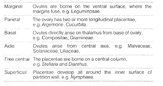
The arrangement of placentae on the ovary wall is
called placentation. The placentation may be

Fruits

After fertilisation of ovary, ovule is changed into seed and
ovary into fruit. The fruit is a characteristic feature of the flowering plants. A true fruit is a ripended ovary. Fruits that
develop from parts other than ovary arc false fruits, e.g. straw berry, apple. The study of fruits is called as pomology.

Classification of Fruits

Fruits can be broadly clas;ified into following three types

(i) Simple Fruits A sinple fruit develops from the single simple (monocarpellay) or compound (polycarpellary)
syncarpous ovary of a flower. In this type, only one fruit formed from one gynoccium. These fruits can be of
following two main types. Dry fruits and fleshy fruits.


(ii) Aggregate (Etaerio) Fruits An aggregate fruit is a group of fruitlcts, w1icb develops from a flower having
polycarpellary apocar·pous (free) gynoecium. The aggregate fruit is also called etaerio, e.g. sadabahar, custard apple, lotus.

(iii) Composite Fruits A composite of multiple fruits develops from the complete inflorescence. These arc of

two types

a) Sorosis A multiple fruit derived from just the pistils of many unisexual flowers of an inflorescence,
e.g. mulberry, jackfruit, pineapple, etc.

b) Syconus A multiple fruit derived from numerous ovaries borne on the inside of the fleshy receptacle of
an inflorescence, e.g. peepal, gular.

Seeds

Seed is a ripened ovule, which contains the embryo. In some seeds, the endosperm (nutritive tissue) is completely consumed
by th developing embryo. Thus, the seeds arc called non-endospermic or exalbuminous seeds, e.g. most dicots
(gram and pea). .

In nnst monocots (maize and rice) and some clicots (castor bean, papaya and cotton) embryo does not consume all
endosperm. So, it persists in the mature seed. Such seeds are called endospermic or albuminous seeds.

PLANTS ANATOMY

'Anatomy' (Gk. Ana-up; tome-cutting) is the study of internal structure of an organisms. Plant anatomy deals with the study
of gross internal structure of plant organs after section cutting of plant parts.

Platnt Tissues

Tissues are the cluster of structurally and functionally similar cells arranged and designed, so as to give the highest possible
efficiency of the function they perform. All cells of a tissue have a common origin .. A tissue may be simple or complex
type. Blood, phloem and muscles arc all examples of tissues.

There are following types of plant tissue

1. Meristematic Tissue

Meristem is a group of cells that have the power of continuous division resulting in the formation of new celL.
These are immature cells. They are present on roots and stems that help them to grow. Meristematic tissues are of
three types based on their position.

(i) Apical meristem Found at the apices of stem and root and function to increase their length. During the
formation of primary plant body, specific regions of this meristem produce dermal tissues, ground tissues
and vascular tissues.

(ii) Intercalary meristem These tissues are intercalated between permanent tissues. It is responsible for
increase in the stem length. Commonly located at the base of the leaves, above the nodes, e.g. grasses or
below the nodes, e.g. mints.

(iii) Lateral meristem Present along the lateral side of stems and roots. Divide in tangential plane, giving ris~
to the secondary permanent tissues to inside and outside and lead to the increase in thickness or girth
of the plant body.

2. Permanent Tissue

These arc formed as a result of division and differcntiatim: in meristcmatic tissues. They lost the power of division.
Permanent tissues are of two types, i.e. simple tissue and complex tissue.


(i) Simple Permanent Tissue A group of similar, permanent cells that performs a
common function is called simple permanent tissue. These are of three types, i.e. parenchyma, collenchyma and
sclcrenchyma.

(a) Parenchyma

They are present in softer parts of plants. This tissue is consist of simple living cells with little specialisation. The
cells are isodiamctric (all sides equal) with thin cell walls (made up of cellulose). Cells are usually loosely packed
with large intercellular spaces. It stores and assimilates food and storage tissue. This tissue provides supports to
plants.

• When the parenchyma cell contains chlorophyll in some situations, it performs photosynthesis. Such type of
parenchyma tissue is called chlorenchyma. It is composed of spongy and mesophyll tissues.


• In aquatic plants, large air cavities are present in parenchyma cells in order to give buoyancy to plants,
which help them to float. Such type of parenchyma tissue is called aerenchyma.

(b) Collenchyma

This is a modified form of parenchyma. These arc the tissues, which are generally found in leaf stalks below the
epidermis, leaf midribs and herbaceous dicot stems.

• Cells are living, elongated and irregularly thickened at the corners. Cell wall has extra deposition of cellulose and pectin and possesses simple pits. They have very little intercellular spaces. The ability to dedifferentiate is nearly absent in collenchyma cells.

• They often contain chloroplasts. It provides mechanical support and elasticity (flexibility) to plants.

• It also allow> easy bending in various parts of a plant (leaf and stem) without breaking.

• They manufacture sugar and starch when possess chloroplasts.


(c) Sclerenchyma
• This type of tissue is present in stems, around vascular bundles, in the veins of leaves and in the hard covering
of seeds and nuts.

The cells of :dcrenchymatous tissue arc dead and do not contain protoplasm.

• The cells arc long and narrow in appearance.

• Cell walls are thickened due to lignin (a chemical substance) deposition, which acts as cement and hardens
them. A prominent middle lamella exists between cells.

• Due to the presence of thick walls, there is no internal space inside the cell.

• It is known to be the chief mechanical tissue, which makes plant r1ards and stiff, e.g. husk of coconut is made
up of sclerenchymatous tissue.

• It provides strength and enables the plant to bear various stresses.

• It forms protective covering around seeds and nuts. It gives rigidity, flexibility and elasticity to the plant body.


(ii) Complex Permanent Tissue


• It is made of more than one type of cells having a common origin. Hence, the cells look different from each
other unlike simple permanent tissue, in which cells are similar in appearance.

• Regardless of different appearances, all the cells coordinate to perform a common function.

• Types of complex permanent tissue are

(a) Xylem (b) Phloem

Both of then are conducting tissues and constitute the vascular bundle. This is a distinctive feature of the
complex plants, which provided them the possibility of
surviving in the terrestrial environment.

(a) Xylem

• It is a vascuhr and mechanical conducting tissue, also known as wood. The cells of xylem have thick walls and
many of them are dead.

• Xylem consists of four types of elements, i.e. tracheids vessels, wood fibre and wood parenchyma.

• It is responsible for the transport of water and minerals from roots to other parts of the plant.

• It also provides mechanical strength to the plant.


(b) Phloem

It is another kind of living conducting tissue, also known as bast.

All phloem cells are living except phloem fibres. Phloem is composed of four types of elements, i.e. sieve tubes,
companion cell, phloem parenchyma and phloem fibres. Sieve tubes are tubular cells with walls perforated by
pores no nucleus. Companion cells are small elongated cell having thin walls.

It transports food from leaves to other parts of the plant.

Materia.ls can move in both directions in it.

Secondary Growth

secondary growth is the growth in the girth of stems and roots in dicots produced by divisions of secondary
meristem, resulting in woody tissue. The cambium is involved in secondary growth. The increase in the
diameter or thickness is due to the formation of Secondary tissues (secondary xylem and phloem) as a
result of the activities of primary and secondary lateral meristems, namely va~;cular cambium (fascicular cambium)
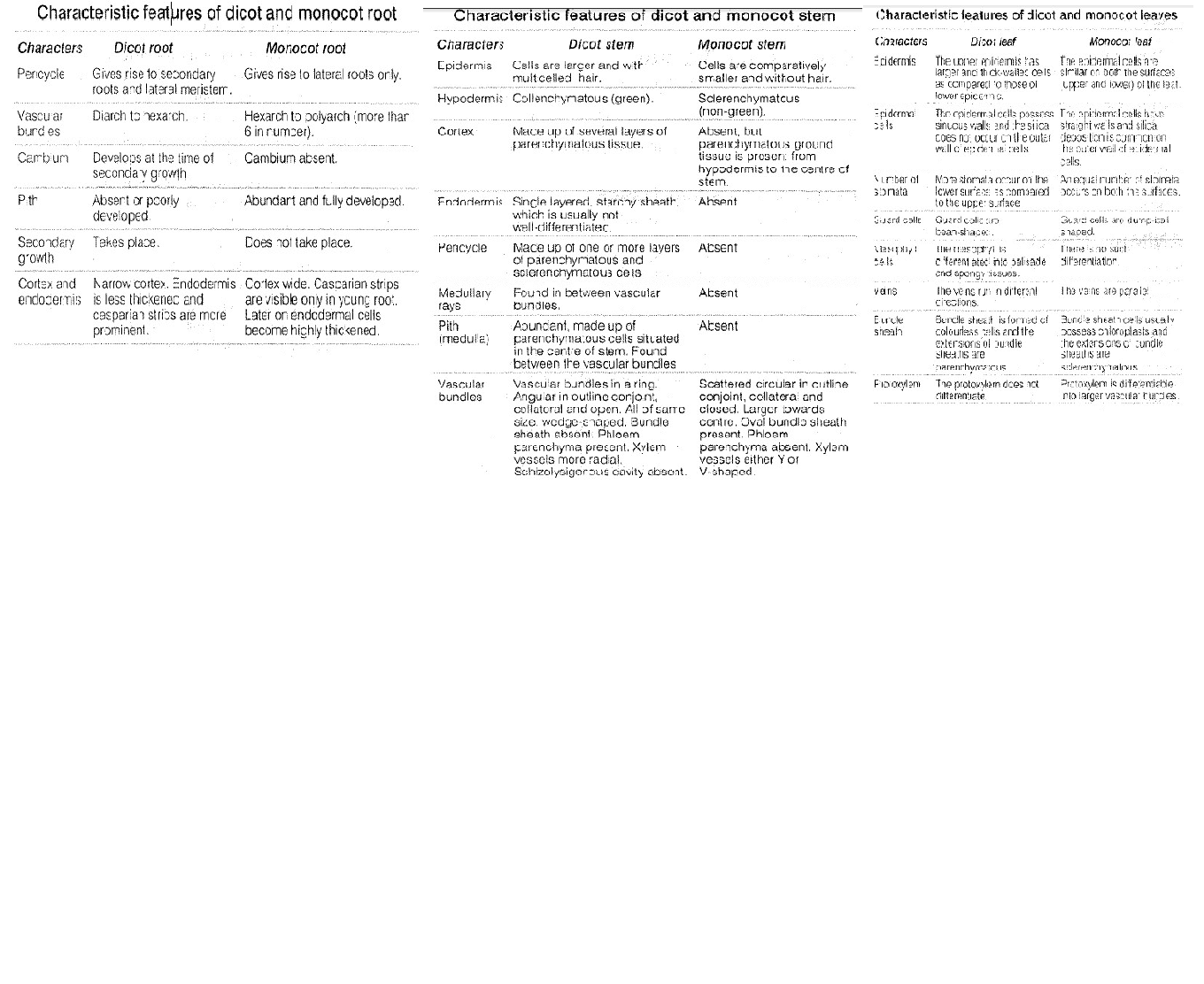
and cork cambium (pellogen), respectively. Secondary growth is seen in only dicot plants. Monocots do not
s1ow secondary growth because cambium is absent in them.

Wood

Wood represents the secondary xylem. It is composed mostly of hollow, elongated, spindle-shaped cells that arc
arranged parallel to each other along the tree trunk. Wood clearly show distinctions, i.e. sapwood (inner wood,
composed of living cdls that conduct sap upward in tree) and heartwood (the outer, less porous dark wood having
c ead cells).

Based on activity in a growth year, wood is of two types

(i) Latewood or Autumn wood It is produced in autumn and have few small xylem clements that
are having small lumen.


(ii) Earlywood or Spring wood It is produced in spring, when cambium is more active and
produces large number of large sized xylem element.


• Thus, each year two zones of secondary xylem are formed, which constitute an annual or growth ring
that determines the age of a plant.

ANIMAL TISSUE

All animals arc structurally organised into cells, tissues, organs or organ systems. The body of all
complex animals consists of four basic types of tissues. A tissue may be defined as a group of similar cells
having a similar origin and specialised for a specific function along with the intercellular substance. The
study of tissues is called Histology. Bichat (Father of Histology) introduced the term 'tissue'. The term
Histology' was coined by Mayer. The tissue arises from the undifferentiated cells of the primary germ
layers (ectoderm, mesoderm and endoderm).

On the basis of their structure and function, animal tissues can be broadly classified into four basic types,
i.e. epithelial, connective, muscular and neural tissues.

These are as follows

1. Epithelial Tissue

The term 'epithelium' (Pl. epithelia) was introduced by Frederik Ruysch in 1703. An epithelium is a tissue
made up of one or more layers of cells, compactly arranged with little intercellular matrix. It covers the
external body surface and lines internal body cavities. Thus, it is also called as covering tissue. Epithelial
tissue also takes part in healing the wounds.

On the basis of cell layers and shape of cells, epithelial tissues arc classified into

(i) Simple Squamous Epithelium It is single layered and closely fitted. It is further
catagorised as

(a) Squamous Epithelium It also covers oesophagus and lining of mouth.

(b) Cuboidal Epithelium It forms lining of kidney tubules and ducts of salivary glands,
where it provides mechanical support. It also forms germinal epithelium of gonads. It also
helps in absorption, excrection and secretion.

(c) Columnar Epithelium It is found in the inner lining of intestine, where absorption
and secretion occur. It facilitates movement across epithelial barrier.

(ii) Stratified Squamous Epithelium Cells arc arranged in many layers and are not similar. It is found in the outer
side of skin as it is highly resistant to mechanical injury and is water-pro. If They are present on body cavity,
cornea of eyes, anus, buccal cavity, etc.

(iii) Pseudostratified Epithelium The epithelium is one-cell thick, but appears two-layered because all the cells do not
reach the free surface. The cells are attached to the basement mcmbrancc, hence they are called
pseudostratified. The mucus secreting goblet cells also occur in this epithelium. It is present in respiratory tract.

2. Connective Tissue

Connective tissue is most abundant, widely distributed body tissue, mesodermal in crigin (with intercellular spaces). Major
functions include binding, support, protection, transport, insulation, fat storage and body defence. They make approximately
30% , part of body. they are broadly categorised into three main types

(i) Proper Connective Tissue

It has a viscous, gel-like matrix composed of proteoglycans. It is of following types

(a) Loose connectiw tissue Cells and fibres are loosely arranged in a semifluid matrix. They are of the following
types


• Areolar tissue It occurs beneath the epithelia of many hollow visceral organs, skin and in blood vessels (arteries and
veins).

• Adipose tissue Locaed mainly beneath the skin, heart, blood vessels, kidney and bone. It is specialised to store fats and
reduces heat loss through the skin. Thus, it keeps the body warm.

There are two types of adipose tissue, white/yellow fat (single layered fat dmplets present in cell surrounded by
small amount of cytoplasm) and brown fat (has multiple small fat droplets svrrounded by larger amount of
cytoplasm).

The former is found in blubber of whales while latter occurs in newborn babies and some
hibernating animals.

(b) Dense connective tisssue It is mainly made up of compactcly packed bundles of collagen fibres
with very little matrix. It is further classified as

• Dense regular co~nective tissue, where collagen fibres are present in rows b~tween
many parallel bundles of fibres. White fibrous, e.g. tendon (connect muscle and bone) and
yellow elastic, e.g. ligament (connect bone and bone) are two of its types.

• Dense irregular connective tissue having fibroblasts with many fibres oriented
differently, e.g. in skin.

(ii) Supportive Connective Tissue

It is of following two types, i.e. cartilage and bone.

(a) Cartilage It is solid, semi-rigid with matrix and is composed of a firm, but flexible material
called chondrin (protein) that is secreted by cells, called chondrocytes. It also contains fibres,
mostly of collagen.

• The cartilage is of three types as following.

• Hyaline cartilage It occurs in the larynx, nasal septum, tracheal rings and costal cartilage and found
at the ends of bones to form articular cartilage. Fibrous cartilage It contains prominent fibres in

matrix of two types.

• White fibrocartilage connects bones like pubis symphysis in pelvis and form intcrvertcrbral discs
and yellow.

• Elastic fibrocartilage It provides strength and maintains shape of car pinna, tip of the nose,
epiglottis, Eustachian tube and larynx.

• Calcified cartilage Cartilage matrix contains granules of calcium carbonate. Found in
suprascapula of pectoral girdle of frog and vertebrae of shark.

(b) Bone It is a solid, rigid connective tissue consisting of four parts, i.e. periosteum, matrix,
endosteum and bone marrow. Completely covered with dense, white fibrous sheath called
pe·riosteum.

• Spaces called lacunae occur in the matrix. Each lacuna is occupied by flat bone cell or osteocyte
(they are rnetabolically inactive cells).

(iii) Vascular Connective Tissue

Also called fluid connective tissue. These are specialised connective tissue that circulate through the
cardeovascular system. It is broadly classified as two main types, i.e. blood and lymph.

Blood

• Blood is a mobile and softest connective tissue. The study of blood is known as haematology. In human beings, volume of
blood is around 5-6 litres. It makes up 6-10% of total body weight.

• Blood makes up the chief transport system in body.

• Blood is salty in taste and it is heavier than water.

• Viscosity of blood is 4.7.

• pH of blood is 7.3 to 7.4, i.e. it is slightly alkaline.

• pH of blood is maintained by balancing the ratio of sodium bicarbonate and carbon:.c acid in blood.

• Buffer of the blood is sodium bicarbonate.

• Oxygenated blood is shining red in colour, whereas deoxygenated blood is pink-purple in colour.

• Acidity of blood results haemoglobin to carry less oxygen.

• pH of blood in arteries is more than in veins.

• People living at higher altitudes, usually have more blood compared to those living at lower altitudes.

'• Blood is made up of two main components

(i) Plasma (ii) Blood cells (blood corpuscles)

Plasma

Plasma represents the matrix of blood, in which blood cells remain embedded.

• Plasma= blood- corpuscles (RBCs + WBCs).

• Plasma is a transparent, slightly alkaline part of blood.

• It forms 55-60%, volume of blood. It contains water (91-92%), solid (8-9%) and inorganic salts (0.9%).

• Solid part of it contains 7% protein (albumin, globulin, fibrinogen, immunoglobulin and prothrombin).

• Albumin protein in plas:na maintains normal blood pressure.

• Similarly, immunoglobulins of blood plasma act as antibodies and help in body defence.


Functions of Blood Plasma

• Retention of fluid in blood.

• Removal of excretory substances.

• Disposal of `CO_2`, transport of `O_2`, distribution of hormones and distribution of vitamins.

• Regulation of water balance.

• It contains antibodies to help resist of infection.

Blood Corpuscles

Blood corpuscles formed in a process called haemopoiesis.

They form upto 40-45%, of blood by volume. These arc mainly of four types, i.e. RBCs, WBCs, platelets and spindle cells.

RBCs (Red Blood Corpuscles)

• RBCs are also known as erythrocytes.

• RBCs of vertebrates are nucleated, whereas those of mammals are non-nud~atcd except camel.

• RBCs of mammals !me nucleus, due to degeneration during development process.


• In the foetus, RBCs are mainly formed in liver and spleen, but after birth they are formed in bone
marrow. Bone marrow is the main site for formation of RBCs.

• Salamander (Amphiuma. means) has largest RBCs (about 80 flm in diametre). Musk deer (Tragulus
javanicus) has the smallest RBCs (1.5 μ m).

• RBCs are biconcave and round in shape.

• Number of RBCs changes due to physiological state.

• Lifespan of RBCs in man is 120 days and number is 5000000/cu mm.
In frog and rabbit their lifespan is 100 and 5C-70 days, respectively.

• Number of RBCs is counted by llaemocytometer.

• Excess of RBCs is known as polycythemia.

• Alone RBC is yellow in color, but it appears red in cluster.

• Excess RBCs are stored in spleen, thus it is also known as blood bank.
Liver is called as the graveyard of RBCs.

• When blood is mixed with distilled water or hypotonic solution, the RBCs increase in volume
and burst. This is known as haemolysis.

• RBCs possess haemoglobin, due to which they appear red in colour. Haemoglobin also acts as
vasculatory respiratory pigment.

• In male (15-16 gm), female (13-14 gm) and in child (16.5 gm) haemoglobin is present in 100 mL of
blood.

• Presence of haemoglobin in blood is measured by Sahli's hacmometer.

• Due to the deficiency of Hb, anaemia occurs. It may be of following types

(i) Pernicious anaemia It is a non-genetic disorder due to the deficiency of vitamin- 12 . In which
number of RBCs decreases and size of RBCs increases, but Hb content is less in RBCs.


(ii) Sickle-cell anaemia It is genetic disorder, in which RBCs become sickle-shaped.

(iii) Thalassemia It is a genetic disorder, in which body does not prepare Hb or RBCs.

(iv) Specticemia It is a sort of blood poisoning.

(v) Nutritional anaemia It is a caused due to the deficiency of iron.

Erythropoiesis

• It is a process of formation of RBCs.

• Stem cells (myeloblast cells or haemocytoblast) are responsible for RBCs formation.

• In man, RBCs formation takes place within 72 hrs.

Functions of RBCs

• Haemoglobin of RBC readily combines with oxygen to form
oxyhacmoglobin.


• In the tissues oxyhacmoglobin readily gives up its oxygen.

Thus, blood transports oxygen to tissues by means of RBCs.

• Maintain pH of blood.

• RBCs also transport `CO_2` .

WBCs (White Blood Corpuscles)

• These are also called as leucocytes.

• These are larger than RBCs and devoid of Hb, so they are colourless.

• Nucleus is present in all WBCs.

• In human blood 8000-9000/cu mm WBCs are present.

• The ratio of RBC/WBC is 600 : 1.

• WBCs play an impo:tant role in defence system, hence called wldier's of body.

• The increase in number of \X!BCs is called as leukemia.

• Abnormally low levd of WBCs is called as leucopenia.

• The movement of WBCs to the site of injury is called as diapedesis.

• The lifespan of WBCs in human is approximate 10-13 days.

W3Cs are of two types

(i) Granulocytes (eosinophils, basophils and ncutrophils).

(ii) Agranulocytes (monocytes and lymphocytes).

Eosinophils (2-8%)

• These cells are also kown as acidophils.

• They arc non-phagocytic.

• Their lifespan is about 10-14 hours.

• They can be stained with eosin dye.

• Their nucleus is usually bilobed.

• The number of eosinophils increases in allergy condition
i.e. asthma and hay fever) and worm infection (e.g. Ascaris).

• They play important role in hypersensitivity.

• These are also important in wounds.

Basophils (2%)

• These are also known as cyanophils.

• :Viinimum number in total WBCs.

• These are phagocytic in nature.

• Their nucleus is usually trilobed.

• Their lifespan is 12-15 days.

• Their number increases in chickenpox.

• They represents mast cells of connective tissue.

• These help in blood coagulation by secreting heparin and histamine.

Neutrophils/Heterophils (65%)

• These arc found in maximum number among WBCs in blood.

• They are phagocytic in nature.

• Their nucleus is multilobed.

• Their number increases in bacterial infection. They are most active type of WBCs

• Their lifespan in blood is 10-12 hours and in tissue is 4-5 days.

Monocytes (6%)

• These are the largest WBCs that are phagocytic in nature.

• Their nucleus is horseshoe-shaped.

• Their lifespan is 28 days.

• They are known as macropoliceman of blood.

• Their number increases in TB (tuberculosis).

• They are produced in lymph glands and spleen.

• They are extremely motile.

• Their lifespan varies from some hours to 1 day.

Lymphocytes (26%)

• These are smallest WBCs.

• Their nucleus is rounded and central.

• Their lifespan is of three days.

• They produce antibodies.

• Their number increases in viral infection.

• These are produced in thymus, spleen and tonsils.

Platelets

• These are found in mammals only.

• They are also known as thrombocytes.

• These are non-nucleated.

• Their size is irregular, oval or spherical.

• Their number is 2-5 lakhs/cu mm.

• These have a lifespan of only one week.

• These are the source of thromboplastin, necessary for blood clotting.

• The blood platelets arc absent from the blood of lower
vertebrates, but possess thrombocytes.

Spindle Cells

• These occur in all vertebrates other than mammals.

• These are like RBCs, but devoid of haemoglobin.

• These are spindle-shaped.

• Their nucleus is spherical or oval and cytoplasm is granular.

• Their main function is similar to that of mammalian blood platelets.

Blood Pressure

• It is the pressure exerted by the flow of blood on the walls of arteries and
measured as millimeter of mercury by the instrument called sphygmomanomett:r.

• It can be felt at certain places in our body viz wrist of the hands, etc.

• It is recorded as systolic/ diastolic.

• It has a high systolic value (normal 120 mm Hg) and low diastolic value
(normal 80 mm Hg1.

• It is lower in the capillaries than in arteries.

• It is usually lower in women than in men.

• Hypertension/High blood pressure Systolic pressure is more than 140 mm Hg and diastolic pressure is more than 90 mm Hg.

• Hypotension/Low blood pressure Systolic pressure is 1: clow 11 0 mm Hg
and diastolic pressure is below 70 rnm Hg.

• The larva of genus chimnomus is called bloodworm. It has haemoglobin, providing red colour to it.

• Uraemia is the prcs<~ncc of more urea in blood.

Blood Glucose

• Usually blood glucose level is about 80-100 mg per 100 mL of blood
12 hours after a normal meal, but its concentration rises soon after a carbohydrate rich ciet.

• If blood glucose level exceeds 180 mg per 100 mL, it starts appearing in
urine, i.e. glycosuria.

• Fasting blood glucose is 70-110 mg/dL, glucose after breakfast (pp) is
110-140.

Blood Cholesterol

• Blood cholesterol in limited amount is important for us, but when it
exceeds it's normal amount, it can be harmful for us. Blood cholesterol
increases due to consumption of excess fats.

• The fats are used in the synthesis of biomembrane, vitamin-D, bile salts
and steroid hormones. Normal blood cholesterol is
80-180 mg in 100 mL of blood plasma. Increased blood cholesterol
may lead to its deposition in the internal wall of blood vessels like
arteries and veins, which causes high blood pressure and heart problems.

• To prevent heart problems, the level of high density lipoproteins (HDL)
should be high and low density lipoproteins (LDL) should be low.

Blood Groups

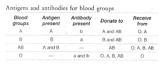
• Karl Landsteiner (Australian pathologist) is known as the father of
blood groups. There are four types of blood groups, A, B, AB and O.

• A, B and O groups were discovered by Landsteiner in 1900, while AB
was discovered by Decastello and Sturle in 1902.

• Blundell discovered the technique of blood transfusion in 1825.

• It is based upon the presence or absence of specific antigens.

• AB blood group is universal recipient (can receive blood from all
blood groups).

• O blood group is universal donor (can donate blood to individuals of
any blood group).

Antigens (Agglutinogens)

• An antigen is stimulus for antibody formation.

• These are present on the surface of RBCs.

• Antigens are proteinaceous in nature.

• A and B are two main antigens.

Antibodies (Agglutins)

These are also known as agglutins.

• Antibodies arc proteins, produced by body in response to the presence of
an antigen.

•These are present in blood plasma. These are produced in lymph nodes and lymph glands.

• These are formed by globulin protein.

• Antibodies are a and b.

Rh factor

• Rh factor is associated with Rh antigen.

• Rh factor was discovered by Landsteiner and Vciner in 1940 in Rhesus monkey.

• Genes, which control Rh factor arc present on autosomes.

• Marriage of Rh+ man and Rh- woman is prohibited because due to this, first birth is safe, while second is
fatal. The disease responsible is known as erythroblastosis foctalis.

• It is because if Rh+ blood is mixed with Rh- blood then antibodies formation starts, i.e. antibodies against
Rh antigen, arc produced in Rh- blood.

• Now-a-days IgG (Immunoglobulin preparation) is given to each Rh- woman after first birth for
prevention of this disease. During blood transfusion Rh factor also plays an important role.

Blood Clotting (Coagulation)

• It is a process of formation of blood clot after inury.

• 3-8 minutes is normal time of blood clot.

• Blood clotting is checked in blood vessels by the presence of anticoagulant (e.g. heparin).

• Anticoagulant removes the cation to check the coagulation.

• Important components of blood clotting are fibrinogen, prothrombin, thromboplastin, calcium ions and
vitamin-K.

• Prothrombin protein of blood clotting is released by liver, while fibrinogen is synthesised in liver.
Haemophilia is a genetic disease, in which blood dotting does not occur.

Functions of blood

(i) Transport of digested food materials (glucose, amino

acids, etc.) and excretory products (`CO_2` ).

(ii) It maintains internal homeostasis.

(iii) Platelets help in blood clotting.

(b) Lymph

It is a colourless fluid connective tissue made up of plasma and WBCs mostly lymphocytes. It is an Extra
Cellular Fluid (ECF), which is intermediate between blood and tissue fluid. It lacks RBCs, platelets and blood
proteins. It carries materials from tissues to blood stream and also in reverse direction. Lymph capillaries present in
the intestinal villi are called lacteals. These are associated with the absorption of digested food.

3. Muscular Tissue

Muscular tissue .s contractile tissue in general, develops from the mesoderm of the embryo.
It consists of long, cylindrical fibres, composed of numerous fine fibrils called myofibrils, which are made of
two proteins called actin and myosin. The presence of these proteins gives striated appearance to the muscle
fibres. The cytoplasm of a muscle fibre is called as sarcoplasm and its endoplasmic reticulum as Sarcoplasmic
Reticulum (SR).


Sarcosomes arc abundant mitochondria present between the myofibrils. Sarcolemma is the plasma membrane of a muscle
fibre, surrounded by basal lamina. These are responsible fo:r movement of body parts (tongue), locomotion, supporting
the bones and other structures. Contractibility, excitability and conductivity are the special features of muscular tissue.
They are categorised into three types


(i) Striated or Striped or Skeletal Muscle

• It is found in hody wall, limbs and also occurs in the tongue, pharynx and oesophagus. These are voluntary
muscles that gets fatigued easily.

• It is cylindrical with unbranched fibres. The cells are multinucleate and bounded by sarcolemma with dark,
anisotropic or A-bands and light, isotropic or I-band~:.

• Each A-band has a light zone Henson's line or H-zonc at its middle, so it is the gap between the actin filaments
extending through myosin filaments.

• Each I-band has a dark membrane, at its centre, the membrane of !Hause or Z-line or Z-band.

• Sarcomere is the part of myofibril between two successive Z-lines (attached on both sides by actin filaments).

• Sarcomere has thick primary myofilaments, composed of protein, myosin and secondary myofilaments composed of
actin, tropomyosin and troponin.

• All bands are nadc of either actin/myosin filaments like A-band (both <.ctin and myosin filan;ents), I-band (actin
filaments) and lJ-band (myosin filaments).


(ii) Non-striated or Smooth Muscle

• These are found in walls of internal organs, such as blood vessels, alimentary canal (also called as visceral muscle).
These are involuntary muscles that do not get fatigued.


• These are spindle-shaped with unbranched fibres that contain a single oval nucleus bounded by plasmalemma
and are compo~:ed of actin and myosin. The length of its fibres is 100-200 μm and diameter is 10 μm. There are no
cross-striations, hence are smooth. They have less extensive SR and less numerous mitochondria.


(iii) Cardiac Muscle

• These are largely confined to the wall of heart. Also, present in the pulmonary veins and superior vena cava.
These arc striated involuntary muscles and never get fatigued.

• These are cylindrical with branched fibres. These are uninudeate, have a rich blood supply and contain actin
and myosin filaments.

• They show cross-striations and contain numerous large mitochondria and glycogen granules.

Functions of Muscles

(i) Heat production Muscles contract and maintain the body temperature in extreme cold.

(ii) Locomotion Muscles help in locomotion by contraction.

(iii) Posture Muscles help in maintaining posture at time of sitting and standing by contraction.

4. Neural/Nervous Tissue

Neurons arc the cells making nervous tissues. Their main function is to receive stimuli and conduct impulses to
control and coordinate body functions. These tissues are devoid of power of division and regeneration. I-Iuman body
consists of approximately 10 11 neurons out of which maximum are present in brain. Neurons are longest cells of
the body. They are functional and structural unit of nervous system.

Structure of Neuron

Structure of neuron consists of following things

• Cell body also ca..led cyton, containing nucleus. Cell body consists of cytoplasm, cell organelles and Nissl's granules.

• Dendrites Nissl bodies, ncurofibrils and mitochondria are present in dendrites. They conduct nerve impulse
towards the cell body and are called afferent processes.

• Axon They conduct nerve impulse away from the cell body.

• It is long, thick, cylindrical structure made of Schwann cells. If axon is covered by myelin sheath then it is
termed as myelinated otherwise non-myelinated. Nodes of Ranvicr arc pbces on axon, where myelin sheath is absent.

Functions of Neuron

• Neurons accept stimuli from the atmosphere and respond accordingly.

• They regulate various other biological activities happen in body.

 
SiteLock