Biology Revision Notes Of Human System - I For NDA

HUMAN SYSTEM - 1

Human body is consists of different organs and organ systems. These organ systems include skeletal system, respiratory system, digestive system, etc. In this chapter we will study about all these systems and how they coordinate with each other, for various functions.

LOCOMOTORY SYSTEM

Locomotory system includes skeletal system and muscular system.

`text(1. Skeletal System)`
Osteology is the study of bones. A human body consists of total 206 bones, while newly born baby have 300 bones. Human skeleton is divided into two parts.
(i) Axial Skeleton It includes the bones of skull, vertebrae, ribs and sternum. It consists of total 80 bones.

(ii) Appendicular Skeleton It consists of total 12() hones. It includes bones of girdles and arms and leg:>. Appendicular system is divided into upper extremity (64 bones) and lower extremity (62 bones).

• Bone is the hardest tissue.
• Ostcoblasts arc bone forming cells.
• Os Penis is a bone found in the penis of rodents. Femur is the longest bone, while stapes (a bone in ear ossicles) is the small est bone.
Fibula is the thinnest bone. Tibia is thin and shining bone.
• Sternum is absent in fishes.
• Tibia fibula is the longest bone of frog.
• Funny bone (ulna) is a bone found in bend of the elbow.
• Spondylitis is the inflammation of vertebrae.
• Arteries of ancient mummies remain intact due to well-preserved fibroblast fibres.
• Synovitis is an inflammation leading to swelling at joint.
• Chondrology is the study of cartilages.
• Fibro cartilage is the strongest cartilage. Skeleton have 12 pairs of ribs in human.
• True ribs are first seven pairs of ribs, while 8th, 9th and
1Oth pairs are false ribs and last two pairs (11th and 12th) are known as floating ribs.
• Cartilage of ear lobes have less blood supply. Tendons and ligaments are dense connective tissues. They are made up of collagen and elastic yellow fibre, respectively. Tendon connects skeletal muscles to bones.
• Ligament attaches one bone to another bone, it is highly flexible and contains very little matrix.
• Bone marrow is absent in birds, but in human, it is the site of production of blood cells in some bones.

Muscular System

Muscles contribute most of the total weight of the body (about 40%).
• Muscles arc helpful in locomotion and movement.
• The study of muscles is called as Myology.
• Muscles have glycogen (polysaccharide), which arc used to obtain energy.
• Muscle tissues are of three types, i.e. striated, non-striated and cardiac. We have studied about muscles in chapter-4.
• Muscles have proteins, which are myoglobin, actin, myosin, etc.
• Muscles of face contract slowly, but can remain contracted for a longer time.
Lactic acid is a chemical, which is responsible for fatigue after muscular activity in human beings.
• Lactic acid accumulates in muscles due to anaerobic respiration.
• Myograph instrument is used to record single muscle twitch.
• Cori's cycle is relationship between muscles glycogen and liver glycogen through lactic acid.
• Motor unit acts as an independent unit of muscle.
• Ca +2 and Mg +2 are required for muscle contraction.
• Myostasis is inflammation of a muscle.
The structure of human skeletal system is as follows

Joints

A joint is the meeting place of two bones. Arthrology is the study of joints.
Joints can be classified into following three types

`text{(i) Immovable joints (Fibrous or Synarthrose Joints)}`

• In these, the adjoining bones arc joined by strong bundles of collagen fibres, so that the bones cannot move freely.
• The bones of the skull articulate with one another in a type of immovable joint called suture, e.g.
(a) Articulation of teeth with mandible and maxillae. Here, peg-shaped roots fit into cone-shaped sockets in the jawbones.
(b) Articulation between the first pair of ribs and the breast bone.

(ii) Slightly Movable (Cartilaginous Joints) In these joints, there is a pad of fibro-cartilage between two adjoining bones, which allows for very slight movements. These are also called symphysis, e.g. joints between pubic of pelvic girdles and the joints between the vertebrae (intervertebral disc).

(iii) Freely Movable (Synovial Joints) In these joints, the movement is possible in one or other directions. These arc of following types

(a) Ball and socket between humerus and glenoid cavity of pectoral girdle, femur and pelvic girdle.
(b) Hinge joint between knee, ankle, elbow, phalanges of digits. ·
(c) Pivot joint between atlas and axis of mammals.
(d) Saddle joint between metacarpal and carpal of thumb.
(c) Gliding joint between radio-ulna and carpals.
(f) Ellipsoidal joint between skull and first vertebra.

RESPIRAATORY SYSTEM

Structurally and functionally the conducting part of human respiratory system is highly complicated. The respiratory system is derived from ernbryonic endoderm. The respiratory system includes respiratory organs and conducting organs. The main respiratory organs of humans are lungs. The lungs and related structures together constitute the respiratory system of humans.

`text(Respiratory Organs)`
• Lungs acts as the major respiratory organs.
• Each lung is covered by double membrane called pleurae.
• In human, the right lung has three lobes and left mg has two lobes.
• The alveoli are the ultimate structural and physiological units of lung.
• Alveoli provide great surface area (like villi of intestine in mammals for gaseous exchange by diffusion.
• Gaseous exchange occurs across the respiratory membrane of the alveoli.
• Residual air of lungs can be traced in alveoli.
• The diaphragm is a dome-shaped muscular structure separating the thoracic and abdominal cavities.
• The diaphragm is characteristic of mammals.

`text(Conducting Organs)`
conducting organs conduct air to lungs. These are as follows
(i) Nostrils Holes of the nose are called nostrils, these are paired openings that open up into the nasal chambers. There are two nasal chambers separated by the nasal septum. Each nasal chamber consists of three regions, i.e. vestibular, tcspiratory and olfactory regions. It consists of many goblet cells.

(ii) Pharynx It provides passage to both air and food.
(iii) Larynx It is the uppermost portion of trachea made up of cartilages in humans. It is also called as voice box. Human larynx consists of glottis, cartilages, hyoid
bone, thyrohyoid membrane, vocal cords, laryngeal ligaments and muscles. Glottis is an opening in the floor of pharynx leading to larynx. There are nine pieces of cartilages (3 are single and 3 are paired).
There is a little difference in the size of the larynx in man and woman. It grows larger and becomes prominent in man, therefore it is called adam's apple in man.
(iv) Trachea The trachea or windpipe is a continuation of the larynx. Trachea is supported by incomplete (C-shaped) rings of hyaline cartilages.
(v) Bronchus These are one pair of small, thin-walled tubular structures formed by the division of trachea at the level of 5th thoracic vertebra.
(vi) Bronchiole The tertiary bronchi is subdivided into smaller branches called bronchioles. Bronchioles can enter into group of alveoli by means of their edges. Each group consists of air sacs.

Respiration

It is the process by which gases at·e exchanged between the blood and the air. It is a physical process. It is simply the intake of oxygen and giving out of carbon dioxide. It includes inspiration and expiration.

`text(Inspiration)`
• It is a process, by which fresh air enters in the lungs.
• In this, diaphragm becomes flat and gets lowered by the contraction of its muscle fibres and increases the volume of thoracic cavity.
• The external intercostal muscles contract and pull the ribs and sternum upwards and outwards direction and thus, the volume of thoradc cavity is increased.

`text(Expirathm)`
• It is a process, by which `CO_2` is expelled out from the lungs.
• Muscle fibres of the diaphragm rt?lax make it convex, decreasing the volume of the \thoracie cavity
• External intercostal muscles relax and pull the ribs downwards and inwards direnion, thus decreeing the size of the thoracic cavity.
• 12-15 times in a minute.
• Newborns breath 40 times per minute,
• Respiration rate is minimum while sleeping.
• The air passage in humans occurs in following matmer

DIGESTIVE SYSTEM

Digestion is the process, in which complex, non-fusible food gets converted into simple fusible food particles with the help of digestive enzymes, e.g. proteins are broken into nmino acids. It consents of following parts.

`text(Alilnentary Canal)`
It is also called gut and s·:arts from mouth to anus. Parts of gut are as follows.

(i) Mouth or Buccal Cavity
It has following parts
(a) Palate forms the rod of mouth and separates mouth from nasal chamber.
(b) Tongue is muscular and also bears test bends to perceive the taste of food.
• It's anterior part is sensitive to sweet taste.
• Saliva secreted from buccal cavity containsOntains enzymes salivary amylase ptyalim) and Lyme which the bacteria).
(c) Teeth arc set in tooth sockets on jawas and are. of types, i.e. incisors (cutting teeth), canies (teanng teeth), premolars and molars (gnndmg teeth).
• An adult has 16 teeth on each jaw.
• In each half of jaws starting from middk to backward there arc incisors-2, tanine-1, molars-3, (2 + 1 + 2 + 3).
• Dental formula in human is 2123/2123.
• A tooth is made up of bone-like materiel called destine.
• A tooth is covered by a white shining material celled enamel.
• Enamel is the hardest part of our body.
• Last molars arc called wisdom. teeth.
• Canines are absent in rabbit and squirrel.
• Milk teeth start erupting after 6 months of birth and appear between 6-24 months.
• Dental formula of milk teeth is 2102/21 02.

`text{(ii) Pharynx}`
• It is small sized (12-14 em), conical part, where food and air passages cross each other.
• It is divided into three parts, i.e. nasopharynx (lies behind the nasal chambers), oropharynx (lies behind the buccal cavity) and laryngopharynx (lowest part of pharynx).

`tex{(iii) Oesophagus}`
• It is a long (22-25 em) narrow, muscular and tubular structure.
• It has longitudinal folds, the oesophageal rugae, which prevent the entry of air in it.
• It conducts the food to stomach by peristalsis.

`text{(iv) Stomach}`
• It acts as a temporary reservoir, which churns the food material. Food after getting churned in stomach is called chyme.
• Stomach secretes gastric juice containing HCI and pepsin (enzyme).
• HCI helps in digestion of food.
• Digestion of protein takes place in stomach and small intestine.
• Stomach mixes food with gastric juice.
• It secretes lipase and amylase in gastric juice.
• Lipase and amylase act on fats and starch (or other sugars), respectively.
• The stomach of ruminant animals is consist of four chambers, i.e. rumen, reticulum, omasum and abomasum.

`text{(v) Small Intestine}`
• It is the longest part (about 2 ft or 6 metre) of
alimentary canal.
• It is divided into three parts, i.e. duodenum (situated
nearest to stomach), jejunum and ileum.
• Duodenum receives the bile-pancreatic duct.
• Inner mucous of small intestine have finger-like projections called villi.
• Villi and micro villi increase the surface area of digestion and absorption of food.
• Pancreatic duct have few enzymes, which act on carbohydrates, fats and proteins.
• Ileum absorbs the fatty acids. Maximum food absorption takes place in ileum only.
• Complete digestion of proteins, fats and carbohydrates occurs in small intestine.

`text{(vi) Large Intestine}`
• It consists of three parts, i.e. caecum, colon and rectum.
• In some herbivores (like horse and ass), caecum is large and is a site of microbial digestion of cellulose.
• In man, caecum is very small vestigial organ and is called appendix. In colon, water is reabsorbed.
• Large intestine docs not secrete enzvyme
• Absorption of food and excess water from undigested food is carried out in large intestine.

Digestive Glands

These secrete digestive juices for the digestion of food. These are of four types.

`text(1. Salivary Gland)`
It produces saliva. Salivary glands secrete salivary amylase
(ptyalin), which converts the starch into maltose. These are of four types
(i) Parotid glands Largest glands present just below the external ear. In these glands, virus causes mumps disease.
(ii) Submaxillary glands These lie beneath the jaw-angles.
(iii) Sublingual glands Smallest glands, which lie beneath the tongue and open at the floor of buccal cavity.
(iv) Zygomatic glands These lie beneath eyes.

`text(2. Gastric Gland)`
It is present in the mucosa of the stomach. These are of three types
(i) Cardiac glands secrete an alkaline mucus.
(ii) Pyloric glands secrete an alkaline mucus.
(iii) Fundic glands each gland has four types of cells.
(a) Peptic/zymogen cells secrete pepsinogen for dry digestion of proteins.
(b) Oxyntic cells secrete HCI.
(c) Goblet cells secrete mucus.
(d) Argentaffin cells secrete gastrin hormone.

`text(3. Liver)`

• It is the largest gland and consists of a large right lobe, a small left lobe and two small lobes called quadrate lobe and caudate lobe behind the main lobes.
• On the right lobe lies gall bladder, which stores bile juice secreted by the liver. Bile juice contains no enzyme, but possesses bile salts and bile pigments (bilirubin-yellow and bilirubin-green).
• Bile juice (acids) are steroid.
• Liver also has some phagocytic cells in between hepatic cells called Kupffer's cells to engulf bacteria and foreign substances. Liver is helpful in blood clotting, formation of glycogen, regulation of blood sugar as reserved food, synthesis ofvitamin-A and storage of some minerals and vitamins.
• Glycogen reserves are utilised in human body for obtaining energy.
• Liver breaks fat to produce cholesterol. It helps in the denomination of amino acids and ammonia into urea.

`text(4. Pancreas)`
It functions both as an exocrine and endocrine gland. It secretes amylase, trypsin and chymotl)'psin, carboxypeptidase, lipase and nucleases in the digestive system. It secretes pancreatic juice, which is helpful in digestion of both starch and proteins.

CIRCULATORY SYSTEM

• Circulatory system was discovered by William Harvey (1628).
• Circulatory system can be either of open type or closed type.
In open circulatory system, blood does not flow in vessels.
• The circulatory system consists of heart and blood vessels.

`text(Heart)`
The heart is a hollow, fibromuscular organ of somewhat conical or pyramidal form with upper broad part, the base and the lower narrow apex, which is slightly directed to the left.

Structure of Heart
• Human heart is situated in the pericardia! cavity.
• The heart wall is made of three layers, i.e. pericardium, myocardium and endothelium.
• Pericardia! fluid is the fluid present between layers of heart wall. It allows frictionless movements of heart and protects it from mechanical shocks.
• In males, weight of heart is about 280-340 g. While in females it is 230-280 g.
• Weight of heart of a newborn baby is 20 g.
• Human heart is consist of two atrium and two ventricles.

`text{(i) Right Auricle or Atrium}`
• It receives deoxygenated blood from superior vena cava and inferior vena cava.
• Right auricle opens in right ventricle through a wide circular right auriculoventricular aperture guarded by tricuspid valve.
• Tricuspid valve regulates unidirectional flow of blood from right auricle to right ventricle.

`text{(ii) Right Ventricle}`

It gives off a large blood vessel called pulmonary artery leading to the lungs, which carries deoxygenated blood to the lungs. Semilunar valve stops blood to return back to ventricle.

`textr{(iii) Left Auricle or Atrium}`
It receives oxygenated blood from lungs through pulmonary vems.
• They have no valve.
• Left auricle opens in left ventricle by an aperture called left auriculoventricular aperture.
• This aperture is guarded by bicuspid valve (mitral valve).
• Bicuspid valve prevents backflow of blood from left auricle to left ventricle.


`text{(iv) Left Ventricle}`
• It gives off a large blood vessels called the aorta.
• Aorta carries oxygenated blood to various body parts.
• The thickest part of the human heart is the wall of the left ventricle.

Circulation of Blood

Blood circulation in humans is an example of double circulation. It includes pulmonary circulation and systemic circulation.
(i) Pulmonary Circulation In this type of circulation, the oxygenated blood is pushed by\ the right ventricle to the lungs for oxygenation through pulmonary artery. The oxygenated blood is brought back to left atrium of heart through pulmonary vein.
(ii) Systemic Circulation In this type of circulation, the oxygenated blood brought to left atrium goes to left ventricle and then passed on to different body parts through aorta. Following diagram shows both pulmonary and systemic circulation.

`text(Cardiac Cycle)`
• The study of structure and functioning of heart is called as cardiology.
• Heart constantly contracts or relaxes, due to which transportation of blood occurs in the whole body.
• Cardiac cycle is the event, during which one heartbeat or one cycle of systole and diastole of cardiac muscle occurs.
• Cardiac cycle consists of systole and diastole of atria and ventricles.
• The cardiac impulse starts from SA node, than via a conducting system, this impulse is conducted to all parts of heart.
• SA node is called as the pacemaker of heart.
• Rate of heartbeat in humans is 72-75 per minute.
• One cardiac cycle takes 0.8 seconds.

`text(Heart Sounds)`
The beating of heart produces sounds, heard by stethoscope. First sound (lubb) is a long and louder sound created by the closure of atrioventricular valves (tricuspid and bicuspid) at the beginning of ventricular systole. Second sound (dupp) is a short and sharper sound created by the closure of semilunar valves towards the end of ventricular systole (beginning of ventricular diastole).

`text(Cardiac Output)`
It is the amount of blood pumped by the heart per minute. Cardiac output = Normal heart rate of an adult per min x Amount of blood pumped by heart per min
`= 72xx 70 = 5040 mL per min (5 Llmin).`

Blood Vessels

In circulatory system, oxygenated and deoxygenated blood is transported to different body parts through different vessels namely arteries, veins and capillaries.

EXCRETORY SYSTEM

The process of elimination of metabolic wastes from the body is called excretion. Removal of excess of water from the body or keep water content constant is called osmoregulation.
Depending on the type of waste, nitrogen elimination is of three types
(i) Ammonotelism is the elimination of nitrogen mainly
in the form of ammonia, e.g. aquatic invertebrates, bony fishes and amphibians.
(ii) Ureotelism is the removal of nitrogen mainly as urea. Ammonia combines with `CO_2` in liver to form urea, e.g. mammals, man, frogs, toads.
(iii) Uricotelism is the elimination of nitrogen mainly as uric acid, e.g. birds, reptiles, many insects. Uric acid is commercially extracted from bird dropping.

`text(Excretory Organs of Invertebrates)`
• Flame cells are the organs of excretion in tapeworm.
• Sponges remmre their wastes through their water canal system.
• In Hydra, cells release waste into coelenteron.
• In annelids, excretion takes place by nephridia.
• In insects, excretion takes place by Malpighian tubules.
• In some arthropods (prawns), green glands also help in excretion.
• Waste material in Amoeba is taken out by plasmalemma.
• Spiders have coxal glands.

Excretory System of Human

Excretory system of human consists of following organs

`text(1. Kidney)`
• Kidney is a paired bean-shaped organ, that is present in abdomen just below the diaphragm.
• The left kidney is placed a little higher than the right kidney (revers in rabbit).
• Concavity of kidney called hilus is always inwardly directed.
• They form urine and drain it ultimately into pelvis of the kidney from where the ureter arises.
• Water reabsorption in kidney is controlled by a hormone called ADH.
• In anuria, kidneys become unable to form urine.

`text(Nephron)`
• It is the functional unit of kidney.
• In one kidney around 1 million nephrons are found.
It has two parts

(i) Bowman's Capsule

(a) It has a network of blood capillaries called glomerulus,. which together form Malpighian body.
(b) Blood enters glomerular capillaries through afferent arteriole and leaves through efferent arteriole.

(ii) Renal Tubule
It consists of three parts
(a) Proximal Convoluted Tubule (PCT).
(b) Loop of Henle with thin descending and thick ascending limb.
(c) Distal Convoluted Tubule (DCT), which opens into collecting duct.
• Skin excretes mainly water and NaCl in sweat. Evaporation of sweat requires latent heat from the body due to which body feels cool. This is known as perspiration.
• Lungs excrete `CO_2` and some water vapour. Liver excretes bile pigments and cholesterol in the bile.

`text(2. Ureters)`

This muscular tube emerges out from the hilum of kidneys. Urine enters the ureter from the renal pelvis.

`text(3. Urinary bladder)`
Sac-like structure, which stores urine temporarily. It is absent in birds.
4. Urethra
• Membranous tube, which conducts urine to the exterior. Urethral sphincters keep the urethra closed except during voiding of urine.
• The act of voiding urine is called micturition.
• An adult man secretes 1-1.8 L of urine in 24 hours.
• The yellow colour of urine is due to the presence of the pigment urochrome. pH of urine should be 5.8.
• When water intake is high, the urine is hypotonic.
• When there is less body water, the urine is hypertonic.

REPRODUCTIVE SYSTEM

A process by which living organisms produce young ones of their own types is called reproduction. It includes asexual and sexual reproduction.
• Asexual reproduction occurs without the formation and union of sex cells, e.g. budding (Hydra), binary fission (Amoeba) and multiple fission (malaria parasite).
• Sexual reproduction includes fusion of male and female gametes.

`text(Male Reproductive System)`

Parts of male reproductive system are as follows Testes
• A pair of glandular, oval-shaped organs present in scrotum.
• Temperature of scrotum is 2°C below the body temperature.
• Endocrine tissues of testes produce testosterone.
• Seminiferous tubules are the structural and functional unit of testes.
• In seminiferous tubules, spermatogenic cells are present,which produce sperm cells. Sertoli cells provide nutrition to sperm cells. Leydig's cells in testes secrete testosterone.

`text(Vas Deferens)`
• Vas deferens is a term used for ducts extending from the epididymis to the ejaculatory ducts.
• These store spermatozoa temporarily and transport them during ejaculation.
• Removal of was deferens is called vasectomy.

`text(Urethra)`
It is a thick-walled muscular duct and it is a common passage for both urine and semen. It is also called urogenital duct. It traverses and opens at the tip of the penis.

`text(Penis)`
It i, erectile copulatory organ. It helps in deposition of sperms into female genital tract.

`text(Accessory Sex Glands)`
Different types of accessory sex glands are as follows.

(i) Seminal Vesicle
• Seminal vesicles (utects masculinus) arc situated behind the urinary bladder.
• About 70% of the semen in seminal fluid is produced by seminal vesicles. Seminal fluid contains fructose, (energy source for sperm) citrate, inositol, prostaglandins and several proteins.
• The product of the testes (spermatozoa) and prostate gland along with the fluid from the seminal vesicle are collectively known as semen. Seminal fluid has a pH of about 7.4.

`text{(ii) Prostate Gland}`
• Prostate is situated around the first part of the urethra.
• Prostate gland secretes a thin, milky fluid that contains calcium, cit rate ion, phosphate ion, a clotting enzyme and a profibrinolysin called a'' semen.
• Enlargement of prostate results in prostatitis, in which urination is difficult or impossible.

`text{(iii) Cowper's Gland}`
It is also known as bulbourethral glands are found in male mammal. The secretion of Cowper's glands act as lubricant for the glans penis. It also neutralises any urine in urethra.

`text(Path of Sperms in Mammals)`

Seminiferous tubules -> Rete testis -> Vas efferentia ->
Epididymis -> Vas deferen -> Urethra -> Vagina

Female Reproductive System

Parts of female reproductive system are as follows

`text(Ovaries)`
Ovaries are primary sex organs present in upper pelvic cavity on both lateral sides of uterus. It produces ova and sex hormone progesterone and oesterogen.

`text{Fallopian Tube (Oviducts)}`
• It extends from ovary to the uterus.
• It conducts egg and provides site for fertilisation.
• Tubectomy is the removal of Fallopian tube.

Uterus
• It is situated above and behind the urinary bladder and
remains attached to the body wall by ligaments.
• The wall of uterus is composed of smooth muscle fibre called myometrium. Lumen of the uterus is lined by a mucous membrane called endometrium.
• It receives ova or egg.
• In werus, foetus gets attached by placenta.

`text(Vagina)`

Accessory Sex Glands

Different types of accessory sex glands are as follows
(i) Bartholin's Gland (Vestibular Gland)
Occurs in female mammals and helps in vestibular lubrication. Bartholin's gland of female corresponds to bulbourethral gland (Cowper's gland) of the male.

(ii) Mammary Glands
The mammary glands are modified sweat glands. Production of milk in mammary glands is stimulated by the hormone protactinium, which is secreted by anterior part of pituitary gland, while the ejection of milk into the ducts of mammary glands is stimulated by the hormone oxytocin, which is secreted by posterior part of pituitary gland.

`text(Gametogemesis)`
• It is the process of formation of gametes in gonads.
• It includes spermatogenesis and oogenesis.
• Spermatogenesis is the formation of sperms. Oogenesis is the formation of eggs (ovum).
• Gametogenesis is controlled by gonadotropic hormones (FSH, LH, ICSH, etc.) secreted by pituitary gland.

`text(Menstrual Cycle)`

• These are cyclic changes occur in the uterus, extending approximately a month period (mesum).
• Menstrual cycle has three phases, i.e. proliferative, secretory and menstrual phase.

`text{(i) Proliferative Phase}`
• FSH stimulates follicle to secrete oestrogen.
• This phase has duration of 10-12 days.
• It is also called follicular phase.

`text{(ii) Secretory Phase}`
• Corpus luteum secretes progesterone.
• This phase has duration of 12-14 days.
• Discharge of ovum from Graafian follicle is known as ovulation. It takes place at 14th day of menstrual cycle under the control of LH.

`text{(iii) Menstrual Phase}`
• If ovum is not fertilised, the corpus luteum degenerates causing sudden fall in the progesterone level.
• Breakdown of endometrium takes place, which results in discharge of blood.
• It is controlled by FSH, LH, oestrogen and progesterone.
• The menstrual cycle and menstruation normally remains suspended during pregnancy and lactation.

`text(Menopause)`
• Ovulation and menstrual cycle are stopped permanently.
• It occurs around 45-50 years of age.
• In this stage, women lose the ability to reproduce.

HUMAN SYSTEM - II

`text(NERVOUS SYSTEM)`
• The neural system is a control system, which coordinates with endocrine system to coordinate various body parts, thus maintaining physiology.
• It gives ability to think and understand.
• Nervous system receives and conducts stimuli to show a specific response against that stimuli. It mainly occurs with the help of neurons.
• Neurons are structural and functional units of nervous system.
• On the basis of function, neurons can be
(i) Sensory (receptor/afferent), which connect sense organs with CNS,
(ii) Motor (effect or/efferent), which connect CNS to defectors like muscles and glands,
(iii) Interreirons/ connector/relaying/adjuster, which occurs in between sensory and motor neurons for distant transmission of impulses.

• On the basis of number of nerve processes, there are four types of neurons namely unipolar neuron (only axon with no drone, e.g. early embryo) bipolar neuron (1 dendrite and 1 axon, e.g. olfactory epithelium and retina), multipolar neuron (1 axon and multiple dendrites, e.g. brain and spinal cord) and pseudounipolar neuron (bipolar but appear as unipolar, e.g. dorsal root ganglion of spinal cord).

• A synapse a gap between two neurons.
• Synapse helps in the transmission of nerve impulse from one neuron to another. The membranes of pre and postsynaptk~ neurons are separated by a synaptic deft.
• Human nervous system is divided into three parts, central nervous system, peripheral nervous system and autonomic nervous system.

1. Central Nervous System (CNS)

CNS is the part of nervous system 1 hat com role whole body and itself. The central nervous system is consist of brain and spinal cord.

`text{(i) Brain}`

It: is the anterior portion of the CNS, which is in
the cranial cavity, cranium of the skull It weight from
1220 to 1400 grams Structurally, it consists of three
membranes (meninges)
• Piamater membrane Innermost thin, very delicate, vascular and inverts the brain closely. ·
• Arachnoid membra.ne outer to piamater thin, webby structure.
• Duramater membrme cutcrmost, tough membrane, adhering cosely tn the inside Brain have following parts.


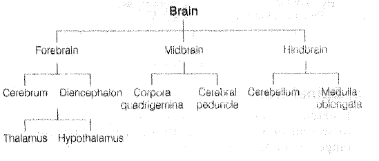
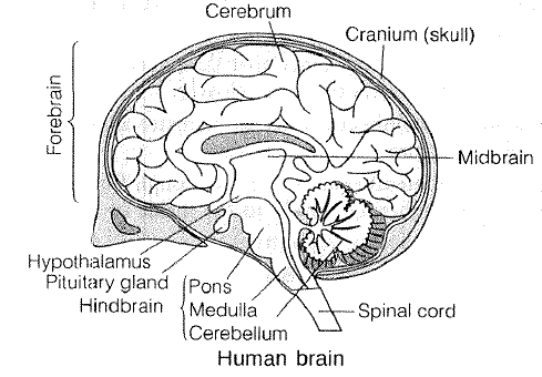
`text{(a) Forebtrain}`
• It is the largest part of brain.
• It occupies 2/3 portion of brain.
• It is the main thinking part of brain.
• It consists of two parts, i.e. cerebrum and diencephalon.
• Cerebrum is the most developed part in human. It is divided into right and left cerebral hemispheres connected by corpus callosum.
• Cerebrum consists of many fold having small grooves (sulci), large grooves (fissures) and bulges between two (gyri).
• Diencephalon is consists of three parts, i.e. thalamus, hypothalamus and epithalamus.
• Thalamus controls emotions and memory.
• Hypotha.lamus control visceral activities. It helps to maintain homeostasis, control thirst, hunger, temperature, respiration, heartbeat.

`text{(b) Midbrain}`
• It is made of two parts, i.e. cerebral peduncle and corpora quadrigema. It lies between the hindbrain and forebrain.
• If consists of group of fibres that arouses the forebrain when something unusual happens.
• Midbrain is responsible for vision and heary.

`text{(c) Hindbrain}`

• It consists of the medulla, cerebellum and pons.
The medulla is the swelling at the tip of the brain that serves as the passage way for nerves extending to and from the brain.
• The pons is the swelling between the medulla and
midbrain. The pons acts as a bridge between various portions of the brain.
• Hind brain is responsible for muscular activities breathing, coughing, etc.

`text{(ii) Spinall Cord}`

• It extends from the base of the brain and is continuous to second lumbar vertebra. In adult, the spinal cord ranges from 42 to 45 em in length.
• It mainly lies in the neural canal of the vertebral column.
• It is basically the posterior part of CNS, which runs mid-dorsally within the vertebral column. The three meninges, i.e. du.ramater, arachnoid and piamater, which covers the brain, also continue over the spinal cord.
• The two indentations, i.e. posterior median calculus and the anterior median tissues separates the spinal cord into left and right haves.
• The inner area is the grey matter, while outside to it are white columns called the white matter.
Functions of Spinal Cord
(i) The stimuli passes from and to the brain through the spinal cord.
(ii) It also act as the centre of spinal reflex action.

2. Peripheral Nervous System (PNS)
• The nerves that originate from central nervous system connect either with receptor or effector organs from peripheral neural system.
• Nerves, which arises from brain are called cranial nerves while the nerves originating from the spinal cord are termed as spinal nerves.
• It relays impulse from the CNS to skeletal muscles. In human body there are 12 pairs of cranial nerves and 31 pairs of spinal nerves.

3. Autonomic: Nervous System (ANS)
It transmit impulse from the CNS to the involuntary organs and smooth mussed of the body. This system was discovered by Langley in 1921. It is further divided into two types
(i) Sympathetic Nervous System Accelerates heartbeat, enlarge pupils, supply blood to muscles, contract nerves of urinary bladder, lowers the intestinal digestion activities, helps in blood clotting, increased secretion of sweat glands, make breathing easier and promote liver to release sugar and decrease bile production are some activities controlled by this nervous system.
(ii) Parasympathetic Nervous System Works just analogs to the sympathetic nervous system, i.e. slows down heartbeat, dilates arteries and lower blood pressure, speeds up peristalsis, stimulate salivary gland secretion, contracts gall bladder, promotes pancreas for secretion, decreases the secretion of sweat glands, etc.

Reflex Action

• A reflex action is an automatic response to a stimulus.
• The pathway taken by nerve impulses in a reflex action is called the reflex arc.
• Reflex actions are under the control of spinal cord (CNS).
• Reflex actions Were discovered by Marshell Hall.
• Some examples of reflex action are moving our foot away when we step on something sharp, moving our hand away on touching a hot plate, a knee-jerk, movement of diaphragm during respiration, coughing, yawning, etc.
Reflex actions arc of two types
(i) Uncondition reflex action
These are rapid actions that are not controlled by brain, e.g. telrs, knee· jerk reflex, etc.
(ii) Conditioned Reflex Action
These actions or activities are learnt by an individual by continuous practice and experience, e.g. dancing, cycling, swimming. These are under the control of cerebral.

SENSE ORGANS

Following four sense organs are found in humans.

`text(Eyes)`
• Eyes arc sense organs for vision.
These have photoreccptors, which convert the energy of specific wavelength of light into action potentials of nerve fibres.
• Eyes are located in the bony cavity called orbit.
• Each eye is a hollow spherical organ often called eyeball.
• The wall of eyeball have three
concentric layers, i.e. sclera, choroid and retina.

`text(Sclera)`
• It is the outer layer made up of white fibrous tissue and having transparent cornea.
• Cornea remain covered by thin and transparent membrane called conjuctiva.

`text(Choroid)`
• Choroid is the middle layer that contains many blood vessels.
• It forms the cilliary body, which inturn form iris.
• Attached to cilliary body a lens is present, which is held in place by ligaments.
• In front of lens pupil is found.
• A transparent watery fluid called aqueous humour fills the space between the lens and cornea.

`textR e t i n a)`

• It is the innermost light sensitive layer.
• It is made up of two types of cells, i.e. photoreceptor rod and cone cells.
• Rods contain a purple coloured photosensitive pigment rhodopsin (formed from vitamin-A) and are sensitive even in dim light and dark. Their function is UV light Vision
Cones have a violet coloured photosensitive pigment iodopsin and are sensitive to bright light and colour perception. The day light vision and colour vision arc functions of light.

• The space between lens and retina is filled with vitreous humour.
• Colour blindness (or Daltonism) is caused due to the deficiency of cones.
• Presbyopia is a visual defect common in older peor I e. In this disease ciliary muscles are weakened causing harcining of lens.
• Myopia (short-sightedness) Image is formed in front of retina. Corrected by using concave lens.
• Hypermetropia (long· sighted ness) Image is formed behind the retina. The person cannot sec the nearby object clearly. The never point of eye gets shifted away from the normal position. It can be corrected by convex lens.

• Many domestic animals and sharks do not possess colour vision.
• Many nocturnal animals like owls have mainly rods in retina and are able to see in darkness. Human eyes are sensitive only to visible range of the spectrum (380-760 nm). ·
• Honeybees can sec ultraviolet light.

Human Ear

Human ear can hear sound of 60-80 decibel. Ear consists of three parts--external, middle and internal ear.
(i) External Ear It consists of an earlobe or pinna and an external auditory canal. It collects and directs soundwaves into the external auditory canal.
(ii) Middle Ear Middle car have three small bones, called ear or auditory ossiclcs. These arc hammer-shaped malleus, anvil-shaped incus and stirrup-like stapes.

(iii) Internal Ear It consists of a bony labyrinth and a membranous labyrinth. The bony labyrinth has three bony semicircular canals, a bony cavity called vestibule and a coiled bony tube called cochlea. It is filled with perilymph.

`text(Tongue)`
• Taste buds arc the organs for taste sensation.
• A taste bud has taste recepor cells, which act as chcmoreccptors. Human tongue possess 10,000 taste buds.
• The anterior part of the tongue is most sensitive to sweet taste, back to the bitter and sides to salty and sour.
• The taste of chillies is a sensation of burning, pain on the pain receptors of the tongue.

`text(Nose)`

• Receptors for smell occur in a modified form of pscudostratified epithelium covering a part of the nasal mucosa. lt is called olfactory epithelium.
• The olfactory receptors cells function as chemoreceptors. They are stimulated by specific chemical substances and produce impulse of smell.

ENDOCRINE SYSTEM

• Endocrine system works in coordination with nervous system.
• It is a system of glands, which pour their secretions, i.e. hormones directly into venous blood or lymph and passage to different body organ in order to control their functioning, metabolism, growth and stress conditions.
• Endocrinology is the branch of Biology, that deals with the study of endocrine glands, hormones and their effects. Thomas Addison (1793-1863) Father of Endocrinology

HORMONES

• These are active messenger (organic) compounds, which are secreted by endocrine cells (glands) of some parts of body and circulates in blood.
• The term hormone was first used by Hippocrates. In 1903, Baylis and Sta ding extracted first hormone from the secretory cells of duodenal mucosa and named it as secretin.
• Hormones arc not found in food. These arc synthesised
in the body itself by endocrine cells. These are mainly three types
(i) Protein hormones, e.g. hormones of parathyroid, pituitary, hypothalamus and pancreas.
(ii) Steroid hormones, e.g. hormones of adrenal cortex and gonads.
(iii) Amino acid derivatives, e.g. hormones of adrenal medulla and thyroid glands like tyrosine derived hormones.

`text(Hormones Vs Enzymes)`
Hormones Vs Enzymes Biological catalysts in living organisms are known as enzymes.
• Besides hormones, enzymes are also compounds of high biological activities, influencing cell metabolism.
`text(Similarities between Hormones and Enzymes)`
•Both are absent in food and are synthesised in cells.
• Both are required in small amounts.
• Role of both in cellular metabolism is specific.
`text(Dissimilarities between Hormones and Enzymes)`
• Hormones arc synthesised only in endocrine cell, while enzymes are synthesised in all cells.
• Hormones do not function outside cells but enzymes can catalyse reactions even outside cells.

Endocrine Glands

Several types of endocrine glands are found in vertebrate body. The location of different glands is given in the figure

`text(Pineal Gland)`
• It is also known as biological clock. It is located on the roof of diencephalon in brain.
• It start degenerating in children at the age of 7 years, in adult it is largely fibrous tissue. It secretes three hormones namely melatonin, ser- tonin and adrcnoglornerulotropin.
• Melationin affects mclanophores.

Pituitary Gland

Pea-shaped endocrine gland present at base of the brain. It is also called master endocrine gland/chief executive gland. It has two parts/lobes
(i) Adenohypophysis (anterior and intermediate lobes)
(ii) Neurohypophysis (posterior lobe)

Hypothalamus

• Hypothalamus secretes both releasing and inhibitory hormones controlling the secretion of some anterior pituitary hormones.
• Appetite, thirst and satiety-centres are located in hypothalamus.

Thyroid

It is largest endocrine gland located in the larynx region of neck. It is bilobed in mammals and birds.
• It secretes thyroxine (1'4 ), tri ido thyronine `(T_3 )` and calcitonin hormone.
• `T_3` and `T_4` increases metabolism, blood pressure and regulate tissue growth.
• Calcitonin regalate calcium levels during childhood.
• Thyroid disorders are more frequent in female than males.
• Hyposecretion in children leads to cretinism and in adults myxocdema (slueggish appearance, feeble mindedness, low body temperature).
• In insufficient quantity of iodine in diet (which is required for the synthesis of thyroxine) is the cause of simple goitre (swelling in the neck).
• Table salt is often iodised for certain area to prevent goitre.
• Excess of thyroxine (hyperthyroidism) leads to increased metabolic activity (highly nervous, overactive and irritated person). Sometimes it leads to exophthalmic goitre or Grave's disease (bulging eyeballs, irregular heartbeat and loss of weight).
• The deficiency of calcitonin results in osteoporosis or loss of bone density.
• Due to age factor or autoimmune disease in thyroid gland, secretion of thyroid reduces, which results in Hashimoto disease.

Parathyroid

• Parathyroid hormone maintains Cal+ level in blood.
• It lowers the serum phosphate.
• It decreases the excretion of calcium.
• Only posecretion Cal+ become less in blood plasma and so uscle contraction decreases and sometimes may stop and may result tetany (parathormone deficiency disease).
• Hypersecretion leads to ostioporosis and weakend bones.

Thymus

• It is an endocrine gland nearest to the heart.
• It is partially endocrine and partially lymphoid gland.
• It produces thymine hormone. It helps in the formation of antibodies in embryo and infants.
• Absence of thymus in embryo brings about lack of lymphocytes, lymph nodes and antibodies.
• T-cells or T -lymphocyt.es are produced in thymus.
• It is responsible for immunity.

Adrenal (Supra Renal) or Emergency Gland

Pancreas

• Exocrine as well as endocrine gland.
• Endocrine part constitutes 2-3%, and represented by 'islets of Langerhans'. The islets have five types of endocrine cells secreting different hormones are

(i) `alpha-cell ->` Glucagon (a hyperglycaemic hormone)
(ii) `beta-cell ->` Insuhin (a hypoglycaemic hormone)
(iii)
(iv) `delta - cells ->` Somatostatin
(v) `F- cells ->` Panceratic polypetide

• Insulin is an anabolic hormone. Testes
• Insulin controls the amount of sugar in blood. Its hyposecretion leads to diabetes mellitus.
• It produces male hormone, i.e. androgens.
• Important androgen of testis is testosterone.
• Glucagon increases blood sugar level.
• Somatostatin controls the functioning of a and `beta -cells.`

Kidney

• It produces erythropoietin hormone, which regulates erythropoiesis or haemopoiesis in bone marrow.
• At puberty by the influence of FSH and ICSH of pituitary, a large amount of testost·rone is produced.
• It also influences ultrafiltration in nephron of kidney,
• It produces female hormone, i.e. oestrogen.
• It also produces renin hormone, which controls aldosterone secretion through renin-angiotensin pathway.

Liver

• It is the largest gland in human.
• It produces angiotensinogen hormone, which changes into angiotensin in the presence of renin of kidney.
• It controls blood pressure.

Gonads

• These z.re reproductive organs in which ova (eggs) and sperms are formed.
• These may also function as endocrine glands, secreting sex hormones. Gonads are of the following types

Testes

• It produces male hormone, i.e. androgens.
• Important androgen of testis is testosterone.
• Testosterone is responsible for deep voice, enlargement of genital organs and appearance of beard in male.
• Testosterone is secreted by Leydig cells.
• Surgical removal of testis is castration or orchidectomy.
• At puberty by the influence of FSH and ICSH of pituitary a large amount of testosterone is produced.

Ovary

• It produces female hormone, i.e. oestrogen.
• Oestrogen is responsible for thin voice, well-developed ovary, oviduct, ma:nmary gland, uterus and vagina in female.
• It helps in controlling of menstrual cycle.
• Corpus luteum is a temporary endocrine gland that produces progesterone hormone.
• It maintains pregnancy.
• It helps in implantation of the embryo or foetus.

 
SiteLock