Biology THE PISTIL, OVULE AND EMBRYO SAC

KEY TOPICS

`star` Pistil
`star` The Megasporangium (Ovule)
`star` Megasporogenesis
`star` Female gametophyte
`star` Nuclear Distribution in Embryo sac
`star` Megasporogenesis at a glance


PISTIL

● The `color{Violet}"Gynoecium"` represents the `color{Violet}"female reproductive part"` of the flower.

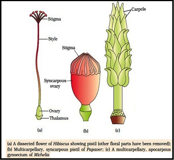
● The gynoecium may consist of a single pistil (`color{Violet}"monocarpellary"`) or may have more than one pistil (`color{Violet}"multicarpellary"`).

● When there are more than one, the pistils may be fused together (`color{Violet}"Syncarpous"`) or may be
free (`color{Violet}"Apocarpous"`).

`star` `color{Brown}"Parts of a Pistil"`:

● Each pistil has three parts, the `color{Violet}"stigma"`, `color{Violet}"style"` and `color{Violet}"ovary"`.

● The stigma serves as a `color{Violet}"landing platform"` for pollen grains.

● The style is the `color{Violet}"elongated slender part"` beneath the stigma.

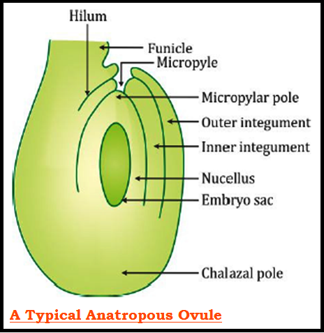
● The `color{Violet}"basal bulged part"` of the pistil is the ovary.

`star` `color{Brown}"Inside of an Ovary"`:

● Inside the ovary is the `color{Violet}"ovarian cavity"` (`color{Violet}"locule"`).

● The `color{Violet}"placenta"` is located inside the ovarian cavity.

● Arising from the placenta are the `color{Violet}"megasporangia"`, commonly called `color{Violet}"ovules"`.

● The number of ovules in an ovary may be `color{Violet}"one"` (wheat, paddy, mango) to `color{Violet}"many"` (papaya, water melon, orchids).

THE MEGASPORANGIUM (OVULE)

● A typical `color{Violet}"angiosperm ovule"` is a small structure attached to the `color{Violet}"placenta"` by means of a stalk called `color{Violet}"funicle"`.

● The body of the ovule `color{Violet}"fuses"` with funicle in the region called `color{Violet}"hilum"`.

● Thus, hilum represents the `color{Violet}"junction"` between ovule and funicle.

● Each ovule has one or two `color{Violet}"protective envelopes"` called `color{Violet}"integuments"`.

● Integuments `color{Violet}"encircle the ovule"` except at the tip where a small `color{Violet}"opening"` called the `color{Violet}"micropyle"` is organised.

● Opposite the micropylar end, is the `color{Violet}"chalaza"`, representing the`color{Violet}"basal part"` of the ovule.

● Enclosed within the integuments is a `color{Violet}"mass of cells"` called the `color{Violet}"nucellus"`.

● Cells of the nucellus have abundant `color{Violet}"reserve food materials"`.

● Located in the nucellus is the `color{Violet}"embryo sac"` or `color{Violet}"female gametophyte"`.

● An ovule generally has a `color{Violet}"single embryo sac"` formed from a megaspore through `color{Violet}"reduction division"`.

MEGASPOROGENESIS

● The process of `color{Violet}"formation of megaspores"` from the megaspore mother cell is called `color{Violet}"Megasporogenesis"`.

● Ovules generally differentiate a `color{Violet}"single"` `color{Violet}"megaspore mother cell (MMC)"` in the micropylar region of the nucellus.

● It is a `color{Violet}"large cell"` containing `color{Violet}"dense cytoplasm"` and a `color{Violet}"prominent nucleus"`.

● The MMC undergoes `color{Violet}"meiotic"` division.

● Meiosis results in the production of `color{Violet}"four megaspores"`.

FEMALE GAMETOPHYTE

● In a majority of flowering plants, `color{Violet}"one"` of the megaspores is `color{Violet}"functional"` while the `color{Violet}"other three degenerate"`.

● Only the `color{Violet}"functional megaspore"` develops into the female gametophyte (`color{Violet}"Embryo sac"`).

● This method of embryo sac formation from a `color{Violet}"single megaspore"` is termed `color{Violet}"monosporic development"`.

● The nucleus of the `color{Violet}"functional megaspore"` divides `color{Violet}"mitotically"` to form two nuclei which move to the `color{Violet}"opposite poles"`, forming the `color{Violet}"2-nucleate"` embryo sac.

● Two more sequential mitotic nuclear divisions result in the formation of the `color{Violet}"4-nucleate"` and later the `color{Violet}"8-nucleate"` stages of the embryo sac.

● These mitotic divisions are strictly `color{Violet}"Free nuclear"`, that is, nuclear divisions are `color{Violet}"not followed immediately"` by cell wall formation.

● `color{Violet}"After"` the 8-nucleate stage, `color{Violet}"cell walls are laid down"` leading to the organisation of the typical `color{Violet}"female gametophyte"` or embryo sac.

NUCLEAR DISTRIBUTION IN EMBRYO SAC

● `color{Violet}"Six of the eight"` nuclei are surrounded by `color{Violet}"cell walls"` and organised into cells.

● The remaining two nuclei, called `color{Violet}"polar nuclei"` are situated below the `color{Violet}"egg apparatus"` in the large `color{Violet}"central cel"`.

● There is a characteristic distribution of the cells within the embryo sac.

`star` `color{Brown}"Egg Apparatus"`:

● `color{Violet}"Three"` cells are grouped together at the `color{Violet}"micropylar"` end and constitute the `color{Violet}"egg apparatus"`.

● The egg apparatus, in turn, consists of `color{Violet}"two synergids"` and `color{Violet}"one egg cell"`.

● The synergids have special `color{Violet}"cellular thickenings"` at the micropylar tip called `color{Violet}"filiform apparatus"`, which play an important role in guiding the `color{Violet}"pollen tubes"` into the synergid.

`star` `color{Brown}"Antipodals"`:

● `color{Violet}"Three"` cells are at the `color{Violet}"chalazal end"` and are called the `color{Violet}"antipodals"`.

`star` `color{Brown}"Central Cell"`:

●The `color{Violet}"large central cell"` , has two `color{Violet}"polar nuclei"`.

`star` Thus, a `color{Brown}"Typical angiosperm"` `color{Brown}"embryo sac"`, `color{Brown}"at maturity"`, `color{Brown}"though 8-nucleate"` `color{Brown}"is 7-celled"`.

MEGASPOROGENESIS AT A GLANCE


SiteLock