Biology CELL ORGANELLES- STRUCTURE AND FUNCTION :

Cytoplasm :

`->` Term "Cytoplasm", was given by Strasburger for the part of cell, presents between the nucleus and cell membrane. Cytoplasm can be devided into two parts .

`->` Ground plasm I Hyaloplasm I Cytosol --> Liquid matrix of cytoplasm except organelles

`->` Trophoplasm -t Part of cytoplasm containing organelles & non living Inclusions.

Cell Organelles :

Permanent Metabolically active and living structures of cytoplasm are called organelles.

Endomembrane System :
`->` While each of the membranous organelles is distinct in terms of its structure and function. many of these are considered together as an endomembrane system because their functions are coordinated.

`->` The endomembrance system include endoplasmic reticulum (ER), golgi complex, lysosomes and vacuoles. Since the functions of the mitochondria, choroplast and peroxisomes are not coordinated with the above components, these are not considered as part of the endornembrane system.
Endoplasmic Reticulum :

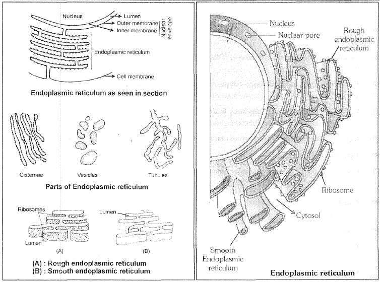
`->` Electron microscopic studies of eukaryotic cells reveal the presence of a network of reticulum of tiny tubular structures scattered in the cytoplasm that is called the endoplasmic reticulum (ER)

Components of E.R. :-

(1) Cisternae- These are long flattened and unbranched units arranged in stacks.

(2) Vesicles- These are oval membrane bound struc1ures.

(3) Tubules - These are irregular, often branched tubes bounded by membrane. Tubules may free or associated with cisternae.

`->` Structure of E.R is like the golgi body but in-E.R. cisternae, vesicles and tubules are isolated in cytoplasm and these do not form complex.

`->` Golgi body is localised cell organelle while E.R is widespread in cytoplasm. E. H. is often termed as "System of Membranes''

`->` ER divide the intracellular space into two distinct cornpalirnent i.e. Luminal (inside ER) and extra luminal (cytoplasm) compartments.

Modifications Of E.R. :

(1) Sarcoplasmic Reticulum (S.R.) :-These smooth E.R. occurs in skeletal and cardiac muscles. S.R Stores `Ca^(+2)` and energy rich compounds required for muscle contraction.

(2) Ergastoplasm :-When the ribosomes are accumulated on the small parallel cisternae of E.R., then called Ergastoplasm:- Ergastoplasm of nerve cells is called as Nissl's bodies.

(3) Myeloid Bodies :-

Myeloid bodies are the specialised smooth E.R which found in pigmented epithelial cells of the retina.

Myeloid body is light sensitive structure and may be involved in pigment migration.

(4) Microsomes - These are pieces of E..R. wilh associated ribosomal pmiicles. These can be obtained by fragementation and high speed centrifugation of cell. They do not exist as such in the living cell. Scientist used microsome for invitro protein synthesis study.

Enzymes Of E.R. :

Sucrases, NADl-1 diphosphatase, Gulcose-6-phosphatase, `NADH` kytochrome-C-reductase, `Mg^(+2)` activated ATPase, Nucleotide diphosphatase, Ascorbic acid synthetase arc enzymes of E.R

Functions Of E.R.

(1) Mechanical support:- Microfilaments, Microtubules and E.R forrns endoskeleton of cell.

(2) Intracellular exchange:- E.R forms intracellular conduc!ing system. Twnsport of materials in cytoplasm from one place to another may occurs through the E. E.

`->` At some places E.H. is also connected to P.M. So E.!-\. can secrete the materials oulside the cell.

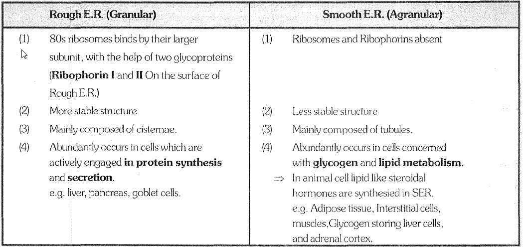
(3) Rough E.R. :-Provides site for the protein synthesis, because rou~Jh LR, has ribosomes on its surface.

(4) Lipid Synthesis:- Lipids (cholesterol & phospholipids) synthesized by the aqranular pmiion of E.R (Smooth E.R.)
The major lipids synthesized by S. E. Rare phospholipids and choleslerol.

(5) Cellular metabolism:-The membranes of the reticulum provides an increased surface for melabolic activities within the cytoplasm.

(6) Formation of nuclear membrane:- Fragmented vesicles of disintegrated nuclear membrane and ER elements arranged around the chromosomes to form a new nuclear membrane during cell division.

(7) Formation of lysosomes, Golgi body & some Micro bodies. All the organelles are form by E.R. which have membrane except chloroplast and mitochondria (semi autonomous organelles)

(8) Detoxification:- Smooth EH concerned with detoxifica1ion of drugs, pollutants and steroids.
`->` Cytochrome `P_450` in E.R. act as enzyme which function in detoxification of dn.tgs
(9) E.R provides the precursor of secretory material to golgii body.

Golgi Complex :

`->` Camillo Golgi (1898) first observed densely stained reticular sltucturc near the nucleus. These were later named Golgi
bodies after him. .

`->` Golgi body also named as.

# Golgibody # Dallon complex # Golgi Complex
# Lipochondria ( rich in lipids) # Baker's bocly # Idiosome
# Dictyosome (plant golgi body) # Trophospongium

#The cytoplasm surrounding Goi~Ji body have fewer or no other organelles. It is called Golgi ground substance or zone of exclusion.

Structure

Golgi complex is made up of four parts -

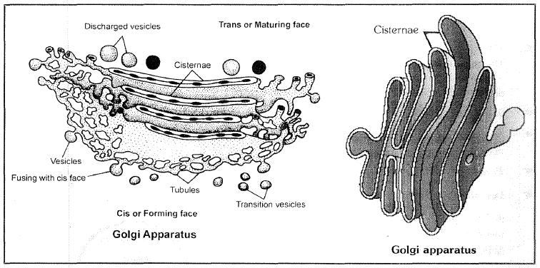
(1) Cisternae:- These are flat disc shaped, sacs like structure many cistenae are arranged in a stack (parallel to each other). Diameter 0.5 pm to 1.0 ~tm. Dense opaque material inside cisternae is called Nodes.

`->` Varied number of cisternae are present in Colgi complex.

`->` Convex surface of cisternae which is towards the nucleus is called cis- face or forming face.

`->` Concave surface of cisternae which is towards the membrane is called Transface or maturing face.

`->` The cis and trans faces of the organelle are entirely different but inner connected.

(2) Tubules:-

These are branched and irregular tube like structures associated with cisternae.

(3) Golgian Vacuoles :-

Large spherical structures associated to tubules.

(4) Vesicles :

Spherical structures arise by budding from tubules. Vesicles are filled with secretory materials.

Functions :

(1) Cell Secretion:- Chief function of golgi body is secretion (export) of macromolecules.

Secretion involve three steps :

(a) Golgi body recieves the materials from E.R. through it's cis- face.

(b) These materials are chemically modilified by golgi body. (For e.g. glycosidation ( glycosylation ) of protiens and lipids takes place in golgi body and it yields glycoprotiens and glycolipids).

(c) A.fter chemical modifications materials are packed in vesicles. These vesicles are pinched off from trans face of golgi body and discharged out side the cell.

Note :

`->` The golg{ apparatus/principally performs the function of packaging materials, to be delivered either to the intra-cellular targets or secreted outside the celL Materials to be packaged in the form of vesicles from the � ER fuse wilth the cis face oflhe golgi apparatus and move towards the maturing face. This explains, why the golgiapparatus remains in close association with the endoplasmic reticulum.

A number of proteins synthesised by ribosomes on the endoplasmic reticulum are modified in the cisternae of the
golgi apparatus before they are released from its trans face. Golgi apparatus is the important site of formation
of glycoproteins and glycolipids.

`->` All the macromolecules which are to be sent out side the cell, move through the golgi body. So golgi body is termed as "Director of macromolecular traffic in cell" or middle men of cell.

(2) Synthesis of cell wall Material (Polysaccharide synthesis)

(3) Cell plate formation (Phragmoplast) during cell formation.

(4) Formation of acrosome during spermiogenesis. (formation of male ~~ametes)

(5) Vitelline membrane of egg is secreted by golgi body.

(6) Formation of Lysosome = It is collective function of golgi body and E.R.

(7) Secretion of hormones by glands

 
SiteLock