Biology MITOCHONDRIA, RIBOSOMES, PLASTIDS, MICROBODIES

RIBOSOMES (ENGINE OF CELL)

`->` Ribosomes are the granular structures first observed under the electron microscope as dense particles by George Palade (1953). They are composed of ribonucleic acid (RNA) and proteins and are not surrounded by any membrane.

`->` Except mammalian RBC all living cells have ribosomes. (Both prokaryotes & Eukaryotes)

`->` Ribosomes are smallest cell organelles (150x250 AD) Eibosomes are organelle without membranes.

`->` Ribosomes are also called as "Organelle within organelle��, "Protein factory of cell", Ribonucleoprotein particle, Palade granule.

Types of Ribosomes :-

(1) Eukaryotic ribosomes :- `80` s- Occur in cytoplasm of eukaryotic cells.

(2) Prokaryotic ribosomes :- `70`s- Occur in cytoplasm and associated with plasma membrane of prokaryotic
cell.

`->` `70`s ribosome also present in mitochondria and chloroplast of eukaryotes. (`55` S ribosome present in mitochondria of mammals)

`->` S=Svedberg unit or Sedimentation rate. It indirectly is a measure of density and size.

`->` Each ribosome composed of two subunits i.e. larger and smaller subunits.

`80`s = `60`s + `40`s

`70`s = `50`s + `30`s


`->` Magnesium ion is essential for the binding the ribosome sub units. `Mg^(+2)` form ionic bond with phosphate groups of r- RNA of two subunits. Minimum `0.001` M `Mg^(+2)` concentration is required for structural formation of ribosomes. If `Mg^(+2)` concentration increased `10` times then ribosome dimer are formed.

`80`s +`80`s = `120`s (Dimer)

`70`s + `70`s = `100`s (Dimer)

Chemical Composition of Ribosomes :

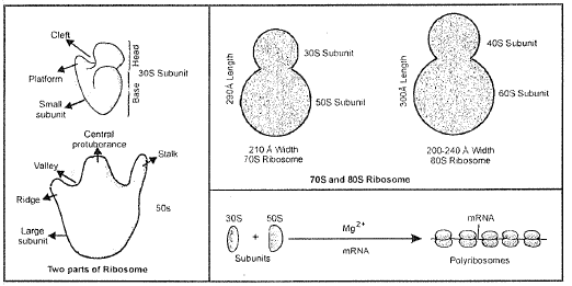
`70`s - `60%` r- RNA + `40%` proteins

`80`s - `40%` r-RNA + `60%` proteins

`60`s - r-RNA `28`s, `5.8`s, `5`s

`40`s - r-RNA `18`s

`50`s - r-RNA `23`s,`5`s

`30`s - r-RNA `16`s

`->` At the time of protein synthesis, several ribosomes become attached to m-RNA with the help of smaller subunits. This structure is called polyribosome or polysome or Ergosome. Ribosomes move along the m-RNA like beads on a string, during protein synthesis. Larger subunit contains peptidyl transferase enzyme (`23`s rRNA) which helps in the formation of peptide bond during protein synthesis. This is an example of Ribozyme. (Noller 1992)

Two sites are found on larger sub units :

(i) A- site -->Acceptor site for l-ENA

(ii) P-site --> site for growing polypeptide chain

After synthesis on ribosomes, protein are transported in cytoplasm and organelles.
The proper folding and transport of proteins is assisted by specific proteins called Chaperons.

CYTOSKELETON

`->` An elaborate network of filamentous proteinaceous structures present in the cytoplasm is collectively referred to as the cytoskeleton. The cytoskeleton in a cell are involved in many functions such as mechanical support, motility, maintenance of the shape of the cell.

MICROTUBULES

`->` Microtubu/es are composed of contractile protien, Tubulin.

`->` In plants microtubules often found associated with cell wall. Probably these transport cell wall material from Golgi body to outside of cell. During cell division these microtubules form spindle fibers.

MICRO FILAMENTS

`->` They are composed of contractile prolien, Actin which concern with muscle contraction, Microtubules and microfilament are part of cytoskeleton-base of cell.

CILIA AND FLAGELLA

`->` Cilia (sing.: cilium) and flagella (sing.: flagellum) are hair-like outgrowths of the cell membrane. Cilia are small structures which work like oars, causing the movement of either the cell or the surrounding fluid. Flagella are comparatively longer and responsible for cell movement. The prokaryotic bacteria also possess flagella but these are structurally different from that of the eukaryolic flagella.

`->` Cilia & Flagella are mechanical, hair like cellular appendages and locomotory structure. Flagellar apparatus is consist of following Parts.

(a) Shaft or ciliary part : It is projecting hair like part of ciliary appartus. Cilium is composed of microtubules. (`9` doublet + `2` singlet)

`->` The electron microscopic study of a cilium or the flagellum show that they are covered cvilh plasma membrane. Their core called the axoneme, possesses a number of microtubules running parallel to the long axis. The axoneme usually has nine doublets of radially cmanged peripheral microtubules, and a pair of centrally located microtubules. Such an arrangement of axonemal microtubules is refen�ed to as the `9+2` array. (`9` double+ `2` singlet)

`->` Arms of A tubules consist of an enzymatic protein dynien similar to myosin of muscle cells. Dynien have ability of hydrolysis of ATP & liberates enerqy for ciliary or flagellar movement.

`->` The central tubules are connected by bridges and is also enclosed by a central sheath, which i.s connected to one of the tubules of each peripheral doublets by radial spoke. Thus there are nine radial spokes. The peripheral doublets are also interconnected by linkers. Both the cilium & flgellum emerge from centriole-like structure called the basal bodies.

(b) Kinetosome or basal granule or Blepheroplast or Basal body:

It is membraneless structure. lies immediately below the plasma membrane. Kasal body exhibil cart wheel structure similar to centriole. (`9` triplet fibriles connected to a central hub).

(c) Rootlet or Rhizoplast :

This is a conical bundle of protein fibers which arises from basal body to differenl directions. Hoollet have dark bands composed of ATPase.

Types of Flagella:

( 1) Whiplash -- When the laterel hair like structures absent.

(2) Tinsel - When the flagella bears lateral hairs like structure (flimmers)

`->` Cilia and Flagella are simialr in structure but some differences may observed

CENTROSOME & CENTRIOLES

`->` Centrosome was discovered by Benden. Boveri named as centrosome. Centrosome is absent in almost all plant cell.

`->` Two centrioles (diplosome) located just outside the nucleus and lie at right angle (`90^0`) to each other. Cytoplasm which surrounds centrioles called as" Centrosphere". Centrioles and centrosphere collectively called centrosome.

Each centriole is surrounded by amorphous peri centriolar mass, which is called as massules or crown or
sateUite.

`->` Centrioles are membraneless elongated structure which exhibit cart wheel structure (just like Basal body of cilia). Basal body is also a type of centriole.

`->` Centriole mainly consist of `9` peripheral triplet fibers of tubulin. (`9 + 0` arrangement)

`->` Centrioles are self duplicating units.

Function :-In animal cells centrioles play important role in initiation of cell division by arranging spindle fibres between two poles of cell. The location of centrioles during cell division decides the plane of division. The plane of division is always at right ang!e to the spindle. Thus centrioles is also termed as "cell centers".

 
SiteLock