Biology CELL CYCLE

�How the cell cycle is controlled

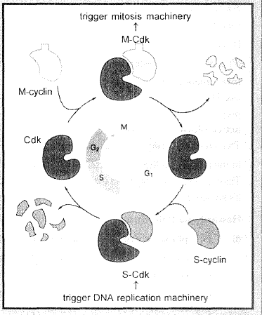
`->` cell cycle is running by a group of special proteins ''CyclinsandCdks(MPF-). (Nurse, TJdunt & .Hartwell 2001. studies.onsaccharomyces (BakerS]east))

`->` A cell reproduces by performing an orderly set sequences of irrevetsible events, In which it duplicates it's contents & then divldes into two; these events are known as cell cyc:le.

`->` Molecular biologists, have made remarkable progress in identifying the biomolecules, that control or drive the cell cycle, many biologists, some of whom worked With invertebrate or frog egg's others with yeast cell or � cell culture. Scientists concluded that the activity of enzymes,. known as cyclin dependant kinases. (Cdk:'s) regulates the cell cycle. Kinase is a.n enzyme that removes a phosphate group from ATP & add to another protein. The kinases involved in the cell cycle are calllef Cdks because they are activated when they combined with key protein called cyclin.

`->` At sorne check points `( tt ( (G_1 -> 5) , (G_2 -> M) ) )` a kinase enzyme combines with cyclin & this moves the cell cycle forwardlly

S-kinase.is capable of starting the replication of DNA after it combined with S-cydin. After cycle forwardly. cyclin is destroyed &.S-kinase is no longer active. M;.kinaseis capable of turning on mitosis after it has bind with M-cyclin . However certain chamcterisiics are universal component of cell cycle controL

2. Division phase

`->` Division phase or M-phase or mitotic phase lasts for only about an hour in the `24` hour duration of cell cycle of a human celL It is the phase of shortest time in cell cycle .

`->` The M-phase represents the phase when the actual cell division or mitosis occurs .

`->` This is the most dramatic period of the cell cycle, involving a major reorganisation of virtually all components of the cell. Since the number of chromosomes in the parent and progeny cells is the same, it is also called as equational division .

`->` Though for convenience mitosis has been divided into four stages of nuclear division, it is very essential to understand that cell division is a progressive process and very clear-cut lines cannot be drawn between various stages .

`->` The M-phae start with nuclear division, corresponding to the separation of daughter chromosome (Karyokinesis) and usually ends with division of cytoplasm (cytokinesis).

`->` Mitosis is divided into the following four stages :-

� Prophase

� Metaphase

� Anaphase

� Telophase

Division of nucleus in mitosis and meiosis is indirect.

(1) Prophase (Longest stage):

`->` Prophase which is the first stage of mitosis follows the `S` and `G_2` phases of interphase .

`->` In the `S` and `G_2` phases the new DNA molecules formed are not distinct but interwined .

`->` Prophase is marked by the initiation of condensation of chromosomal material. The chromosomal material becomes untangled during the process of
chromatin condensation. Chromatin threads
condenses to form chromosomes.

`->` Metabolism of cell decreases, cytoplasm becomes viscous, refractive and pale .

`->` The centriole, which had undergone duplication during S phase of interphase. now begins iG move opposite poles of the cell.

`->` Astral ray forms due to gelation of proteins around centrioles. Spireme stage of chromosome .

`->` Anastral and Amphiastral Mitosis : In higher plants, centrioles are absent and no asters are without asters is known as anastral mitosis. In animals, the asters are present and the mitosis is described as amphiastral, or astral mitosis.

`->` The completion of prophase can thus be marked by the following characteristic events:

� Chromosomal material condenses to form compact mitotic chromosomes. Chromosomes are composed of two chromatids attached together at the centromere.

� Initiation of the assembly of mitotic spindle, the microlubules, the proteinaceous cytoplasm help in the process.

� Cell at the end of prophase when viewed under the microscope, do not show golgi complexes, endoplasmic reticulum, nucleolus and nuclear envolope.

(2) Metaphase :

`->` The complete disintegration of the nuclear envelope marks the start of the second phase of mitosis, hence the chromosomes are spread through the cytoplasm of the cell.

`->` By this stage, condensation of chromosomes is completed and they can be observed clearly under the microscope. This then, is the stage at which morphology of chromosomes is most easily studied.

`->` At this stage, metaphase chromosome is made up of two sister chromatids, which are held
together by the centromere. Small disc-shaped structures at the surface of the centromeres are called
kinetochores. These structures serve as the sites of attachment of spindle fibres (formed by the microtubules) to the chromosomes that are moved into position at the centre of the cell

`->` Spindle fibres attach to kinetochores of chromosomes.


`->` Hence, the metaphase is characterised by all the chromosomes coming to lie at the equator with one chromatid of each chromosome connected by its kinetochore to spindle fibres from one pole and its sister chromatid connected by its kinetochore to spindle fibres from the opposite pole. The plane of alignment of the chromosomes at metaphase is referred to as the metaphase plate.

`->` Chromosomal fibres (discontinous which run from pole to centromere) and supporting fibres (continous which run from pole to pole) arrange in cell.

`->` Spindle fibre are composed of `97%` tubulin protein and `3%` RNA.

`->` Centromere lies at equator and arms remain directed towards poles.

`->` Chromosomal fibres have polarity i.e. + end at equator and end at the pole.

`->` Two chromatids of a chromosome repulse each other and the arms of chromosomes are directed towards the opposite poles. Condensation of chromosomes is completed.

`->` The key features of metaphase are:

� Chromosomes are moved to spindle equator and get aligned along metaphase plate through fibrer to both poles.

(3) Anaphase :(Shortest stage)

`->` In early anaphase interzonal fibres (small and contracted) appears at equator of cell.

`->` Centromere of each chromosome splits lengthwise (division of centromere). Sister chromatids separate from each other and new each chromatid reffered to as individual chromosome.

`->` Number of chromosome becomes double in cell during mitotic anaphase.

`->` Interzonal fibres expands and they push chromosomes towards the opposite poles. (Pushing)

`->` Chromosomal fibres contract and they pull chromosome towards opposite poles. (Pulling)

`->` By pulling and pushing mechanism chromosomes rapidly move towards the opposite poles.

`->` As each chromosome moves away from the equatorial plate, the centromere of each chromosome is towards the pole and hence at the leading edge, with the arms of the chromosome trailing behind .

`->` Approximately 30 ATP are required to carry a chromosome to pole. Chromosomes reach at poles in late anaphase.

Anaphase stage is characterised by the following key events:

� Centromeres split and chromatids separate .

� Chromosome with one chromatids move to opposite poles .

(4) Telophase (Reverse prophase):

At the beginning of the final stage of mitosis, i.e., telophase, the chromosomes that have reached their respective poles decondense and lose their individuality. The individual chromosomes can no longer be seen and chromatin material tends to collect in a mass in the two poles. This is the stage which shows the following key events:

� Chromosomes cluster at opposite spindle poles and their identity is lost as discrete elements.

� Nuclear envelope assembles around the chromosome clusters.

� Nucleolus, golgi complex and ER reform.

 
SiteLock