The Eye
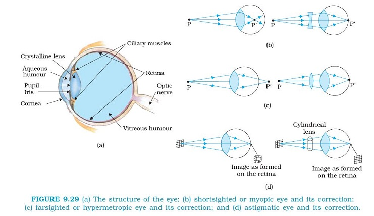
Figure (a) shows the eye. Light enters the eye through a curved front surface, the cornea. It passes through the pupil which is the central hole in the iris. The size of the pupil can change under control of muscles. The light is further focused by the eye lens on the retina. The retina is a film of nerve fibres covering the curved back surface of the eye. The retina contains rods and cones which sense light intensity and colour, respectively, and transmit electrical signals via the optic nerve to the brain which finally processes this information. The shape (curvature) and therefore the focal length of the lens can be modified somewhat by the ciliary muscles. For example, when the muscle is relaxed, the focal length is about 2.5 cm and objects at infinity are in sharp focus on the retina. When the object is brought closer to the eye, in order to maintain the same image-lens distance (≅ 2.5 cm), the focal length of the eye lens becomes shorter by the action of the ciliary muscles. This property of the eye is called `text(accommodation)`.
If the object is too close to the eye, the lens cannot curve enough to focus the image on to the retina, and the image is blurred. The closest distance for which the lens can focus light on the retina is called the least distance of distinct vision, or the near point. The standard value for normal vision is taken as 25 cm. (Often the near point is given the symbol D.) This distance increases with age, because of the decreasing effectiveness of the ciliary muscle and the loss of flexibility of the lens. The near point may be as close as about 7 to 8 cm in a child ten years of age, and may increase to as much as 200 cm at 60 years of age. Thus, if an elderly person tries to read a book at about 25 cm from the eye, the image appears blurred. This condition (defect of the eye) is called `text(presbyopia)`. It is corrected by using a converging lens for reading.
Thus, our eyes are marvelous organs that have the capability to interpret incoming electromagnetic waves as images through a complex process. In spite of all precautions and proactive action, our eyes may develop some defects due to various reasons. We shall restrict our discussion to some common optical defects of the eye. For example, the light from a distant object arriving at the eye-lens may get converged at a point in front of the retina. This type of defect is called `text(nearsightedness)` or `text(myopia)`. This means that the eye is producing too much convergence in the incident beam. To compensate this, we interpose a concave lens between the eye and the object, with the diverging effect desired to get the image focused on the retina [Fig. (b)].
Similarly, if the eye-lens focuses the incoming light at a point behind the retina, a convergent lens is needed to compensate for the defect in vision. This defect is called `text(farsightedness)` or `text(hypermetropia)` [Fig. (c)].
Another common defect of vision is called `text(astigmatism)`. This occurs when the cornea is not spherical in shape. For example, the cornea could have a larger curvature in the vertical plane than in the horizontal plane or vice-versa. If a person with such a defect in eye-lens looks at a wire mesh or a grid of lines, focusing in either the vertical or the horizontal plane may not be as sharp as in the other plane. Astigmatism results in lines in one direction being well focused while those in a perpendicular direction may appear distorted [Fig (d)]. Astigmatism can be corrected by using a cylindrical lens of desired radius of curvature with an appropriately directed axis. This defect can occur along with myopia or hypermetropia.