MIcroscopic Anatomy Of Testis & Ovary
1. TESTES :
The testes are the primary sex organs. They are about 4 � 5 cm long, 2.5 cm wide and 3 cm thick. They are suspended in the scrotal sacs by spermatic cords.
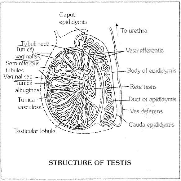
(1) Protective coats (Tunicae) : Each testis has three coverings � tunica vaginalis, tunica albuginea and tunica vasculosa. On one side each testis is covered by hollowed tunica vaginalis, a bilayer of peritoneum with a narrow coelomic cavity having coelomic fluid for sliding. The actual covering of testis is a fibrous connective tissue sheath called tunica albuginea. Tunica albuginea also projects inside testis to form a vertical coloumn called mediastinum and a number of transverse septa.
(2) Testicular lobules : In growth of the tunica albuginea, called septa, divide the testis into some 200 to 300 lobules. Each testicular lobule contains 1 � 4 highly convoluted seminiferous tubules, blood vessels and nerve embedded in loose connective tissue. A total of about 1000 seminiferous tubules occur in each testis. Each tubule is about 70 � 80 cm long. In seminiferous tubules lumen develop after puberty. The ends of the seminiferous tubules converge toward the middle of the posterior surface of the testis and join to form short straight tubules called tubuli recti. The tubuli recti open into a network of wider, irregular tubules called rete testis. Here some of the epithelial cells bear a single cilium to aid sperm transport.
Seminiferous tubules : Each seminiferous tubules is lined by germinal epithelium, seminiferous tubules is the site of spermatogenesis. The process occurs in waves along the length of the tubule, taking about 9 weeks (63 days) to complete in man. Seminiferous tubules contain 2 types of cells �
(i) Germ cells : These are spermatogenic cells by mitotic divisions, produce spermatogonia into the lumen of the seminiferous tubule. The spermatogonia grow into primary spermatocytes which undergo meiosis, producing haploid cells, first secondary spermatocytes and then spermatids. Spermatids differentiate by a process of spermiogenesis into dimorphic haploid sperm (containing X or Y chromosome). Mature spermatozoa lie free in the cavity of the seminiferous tubules.
(ii) Somatic cells / Sertoli cells / Sustentacular cells / Nurse cells : These are supportive nutritive and secrete a polypeptide hormone called inhibin and a steroid estradiol which interferes with spermatogenic activity and kinetics of sperm production.
These are scattered irregularly between spermatogonia.
They also phagocytose damaged germ cells and secrete enzymes for sperm maturation.
Rest on the basement membrane of the seminiferous tubule and its cytoplasm fills all the narrow spaces between the cells of the spermatogenic series. They have ovoid nucleus; exhibits deep identation and has large nucleolus. It has mitochondria, rough endoplasmic reticulum, and lipid droplets.
Sertoli cells is the characteristics of mammalian testis.
Sertoli cell acts as 'Nurse cell' and provide mechanical and metabolic support to developing germ cells.
These cells mediate some important regulatory processes. Sertoli cells produce androgen binding protein (ABP) which serves as vector for androgen and thus generate a hormonal milieu synergistically with FSH to facilitate spermiogenesis.
They do not divide and thus their number is constant.
They are resistant to exogenous / endogenous challenges.
The testes are the primary sex organs. They are about 4 � 5 cm long, 2.5 cm wide and 3 cm thick. They are suspended in the scrotal sacs by spermatic cords.
(1) Protective coats (Tunicae) : Each testis has three coverings � tunica vaginalis, tunica albuginea and tunica vasculosa. On one side each testis is covered by hollowed tunica vaginalis, a bilayer of peritoneum with a narrow coelomic cavity having coelomic fluid for sliding. The actual covering of testis is a fibrous connective tissue sheath called tunica albuginea. Tunica albuginea also projects inside testis to form a vertical coloumn called mediastinum and a number of transverse septa.
(2) Testicular lobules : In growth of the tunica albuginea, called septa, divide the testis into some 200 to 300 lobules. Each testicular lobule contains 1 � 4 highly convoluted seminiferous tubules, blood vessels and nerve embedded in loose connective tissue. A total of about 1000 seminiferous tubules occur in each testis. Each tubule is about 70 � 80 cm long. In seminiferous tubules lumen develop after puberty. The ends of the seminiferous tubules converge toward the middle of the posterior surface of the testis and join to form short straight tubules called tubuli recti. The tubuli recti open into a network of wider, irregular tubules called rete testis. Here some of the epithelial cells bear a single cilium to aid sperm transport.
Seminiferous tubules : Each seminiferous tubules is lined by germinal epithelium, seminiferous tubules is the site of spermatogenesis. The process occurs in waves along the length of the tubule, taking about 9 weeks (63 days) to complete in man. Seminiferous tubules contain 2 types of cells �
(i) Germ cells : These are spermatogenic cells by mitotic divisions, produce spermatogonia into the lumen of the seminiferous tubule. The spermatogonia grow into primary spermatocytes which undergo meiosis, producing haploid cells, first secondary spermatocytes and then spermatids. Spermatids differentiate by a process of spermiogenesis into dimorphic haploid sperm (containing X or Y chromosome). Mature spermatozoa lie free in the cavity of the seminiferous tubules.
(ii) Somatic cells / Sertoli cells / Sustentacular cells / Nurse cells : These are supportive nutritive and secrete a polypeptide hormone called inhibin and a steroid estradiol which interferes with spermatogenic activity and kinetics of sperm production.
These are scattered irregularly between spermatogonia.
They also phagocytose damaged germ cells and secrete enzymes for sperm maturation.
Rest on the basement membrane of the seminiferous tubule and its cytoplasm fills all the narrow spaces between the cells of the spermatogenic series. They have ovoid nucleus; exhibits deep identation and has large nucleolus. It has mitochondria, rough endoplasmic reticulum, and lipid droplets.
Sertoli cells is the characteristics of mammalian testis.
Sertoli cell acts as 'Nurse cell' and provide mechanical and metabolic support to developing germ cells.
These cells mediate some important regulatory processes. Sertoli cells produce androgen binding protein (ABP) which serves as vector for androgen and thus generate a hormonal milieu synergistically with FSH to facilitate spermiogenesis.
They do not divide and thus their number is constant.
They are resistant to exogenous / endogenous challenges.