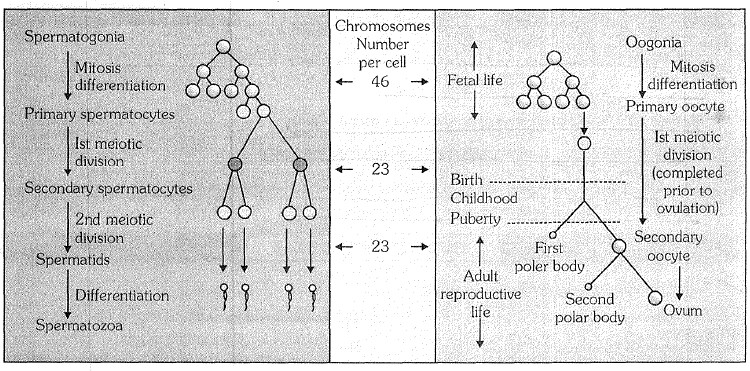
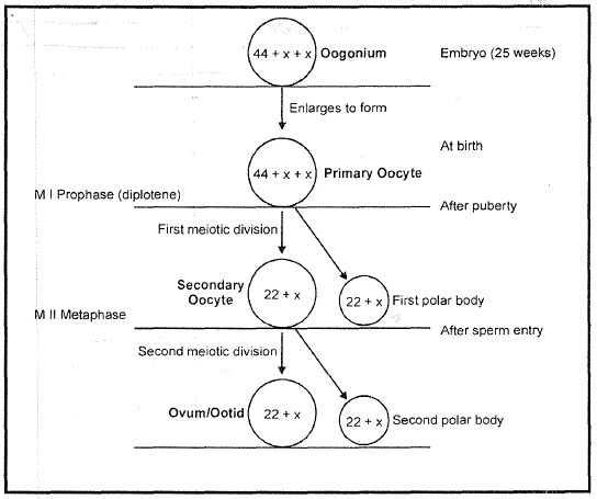
Oogenesis takes place in the ovaries. Unlike sperm formation that starts at puberty, egg formation begins before birth but is completed only after fertilization. Oogenesis consists of three phases �
(a) Multiplication phase
During foetal development, certain cells in the germinal epithelium of the ovary are larger than others and also have larger nuclei.
These cells undergo mitotic divisions, producing undifferentiated germ cells called oogonia or egg mother cells in the ovary.
The oogonia have diploid, number of chromosome, 46 in humans.
The oogonia multiply by mitotic divisions and produce ovigerous cords or egg tubes of pfluger in mammals.
(b) Growth phase : It is prolonged and slow. Oogonia form rounded masses or egg nests at the tips of egg tubes of pfluger.
An egg nest forms ovarian follicle (Graffian follicle) one central oogonium grows and functions as primary oocyte. The others form the covering follicular cells. the latter provide nourishment to primary oocyte. Some nourishment also comes from outside. Yolk is deposited in this state. This phenomenon is called vetellogenesis.
In cooperation with follicular cells, the enlarged primary oocyte secrete mucoprotein membrane or zona pellucida outside its own plasma membrane or vitelline membrane. There is increase in reserve food, size of nucleus, number of mitochondria; functioning of golgi apparatus and complexing of endoplasmic reticulam.
(c) Maturation phase
Meosis occurs. Nucleus shifts towards animal pole and undergoes meosis � I. A daughter nucleus alongwith small quantity of cytoplasm is extruded as primary polar body or polocyte below zona pellucida. Simultaneously primary oocyte is changed into haploid secondary oocyte. It proceeds with meosis � II but stops at metaphase-II. Ovum is generally shed in secondary oocyte stage.
After fertilization, the second meotic division is completed with unequal cytoplasmic cleavage. This forms a large cell the ootid with essentially whole of the cytoplasm, and a very small cell, the second polar body. The ootid and the second polar body are haploid as the second meotic division is equational. The first polar body may divide at about the same time into two polar bodies.
One primary oocyte forms, after two meiotic division, one haploid ootid and three haploid polar bodies. The ootid grows into a functional haploid ovum.
The polar bodies have no function and disintegrate due to lack of cytoplasm and food.
The formation of non functional polar bodies enables the egg to get rid of excess chromosomes. The unequal cytoplasmic division enables the ovum to retain the whole of cytoplasm of the primary oocyte in it for the development of the future embryo.
In humans, ova are released from the ovary in the secondary oocyte stage. Their maturation is completed in the mother's genital tract, usually after the sperm has entered for fertilization.
(iv) Structure of ovum : An ovum is generally spherical, nonmotile gamete with yolky cytoplasm and enclosed in one or more egg envelops. Size of ovum varies in different animals and depends upon the amount of yolk. Size of ovum varies from 10 to a few cm. Largest sized egg is of ostrich and is about 170*135 mm. Egg size and yolk amount are interdependent. It is about 50micron in many polychaete worms, 150micron in tunicates but very large sized in birds and reptiles. In mammals, it is generally microlecithal and about 100micron.
Human ovum is microlecithal with large amount of cytoplasm. Cytoplasm is differentiated into outer, smaller and transparent exoplasm or egg cortex and inner, larger and opaque endoplasm or ooplasm.
Egg cortex is with some cytoskeletal structures like microtubules and microfilaments (Balinsky, 1981), pigment granules and cortical granules of mucopolysaccharides. Endoplasm is with cell-organelles, informosomes tRNAs, histones, enzymes etc. Nucleus of ovum is large, bloated with nucleoplasm and is called germinal vesicle. Nucleus is excentric in position so human ovum has a polarity. The side of ovum with nucleus and polar body is called animal pole, while the opposite side is called vegetal pole.
Egg envelopes. Human ovum is surrounded by a number of egg envelopes :
(a) Vitelline membrane : It is inner, thin, transparent and is secreted by ovum itself.
(b) Zona pellucida : It is middle, thick, transparent and non-cellular.
(c) Corona radiata : It is outer, thicker coat formed of radially elongated follicular cells. Between the vitelline membrane and zona pellucida, there is a narrow perivitelline space.