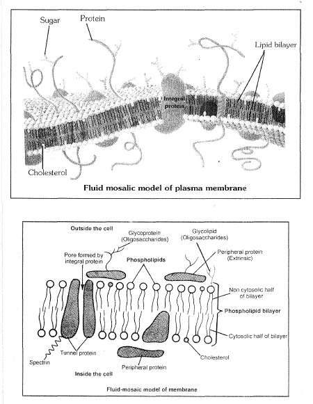
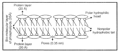
Cell Envelope
Cell envelope is made up of two main components-
1. Cell Wall
2. Cell Membrane
#CELL WALL
(1) Discovery : It was first discovered by Robert Hooke in 1665.
Cell wall is the outer most, rigid, protective, non living and supportive layer found in all the plant cells, bacteria, cyanobacteria and some protists. It is not found in animal cells.
(2) Chemical composition : Mainly cell wall consists of two parts, matrix and cellulosic fibres (microfibriles). Matrix consists of hemicellulose, pectin, glycoproteins, lipids and water. A cellulose molecule is long unbranched chain of glucose molecules. There are about 6,000 glucose units in each chain. In most of the plants cell wall is made up of cellulose a polymer made-up of unbranched chain of glucose molecule linked by glycosidic bond. About 100 molecules of cellulose form a micelle, about 20 micelle form a microfibril and approx 200 microfibril form a fibril. The cell wall of bacteria and the inner layer of blue green algae is made-up of mucopeptide and not of cellulose. The mucopeptide is a polymer of two amino sugars namely N-acetyl glucosamine (NAG) and N-acetyl muramic acid (NAM) held alternately in �1,4- linkage. In higher fungi, the cell wall is made up of chitin, polymer of glucosamine.
Pectin is a mixture of polymerised and methylated galacturans, galacturonic acid and neutral sugars. Hemicellulose is a mixture of polymerised xylans, mannans, glucomannans, galactans, xyloglucans and arabinogalactans. Glycoproteins are known to influence metabolic activities of the wall. A glycoprotein called extensin or expansin takes part in loosening and expansion of cell was through incorporation of cellulose molecules to cellulose microfibrils.
Plant cell wall may have lignin for strength (e.g., woody tissue), silica for stiffness and protection (e.g., epidermal cells of grasses, Equisetum), cutin for preventing loss of water (e.g., epidermal cells), wax as component of cuticle and surface bloom as water repellent (floating leaves) and checking transpiration, suberin for impermeability (e.g., cork cells, endodermal cells), etc.
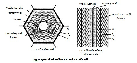
(3) Structure : Cell wall consists of middle lamella, primary wall, secondary wall, tertiary wall.
(i) Middle lamella : Middle lamella is the outermost region which functions as a cementing layer between two cells. It is absent on the outer free surface. It ruptures to create intercellular spaces. Middle lamella is formed of calcium and magnecium pectate. Fruit softening is due to gelatinisation of pectic compounds of middle lamella. Pectin is used as commercial jellying agent. Which is present outside the primary wall.
(ii) Primary wall : A young plant cell forms a single layer of wall material. This layer is known as the primary cell wall. The primary wall is thin, elastic and capable of expansion in a growing cell. It grows by intussusception. Meristematic and parenchymatous cells have primary cell wall only. The cells of leaves and fruits too have only primary wall.
(iii) Secondary wall : In mature cell, more layers of wall material are added internal to the primary wall. These are called the secondary cell wall. Growth by addition of new wall material on the primary wall is called accretion. The secondary wall is thick and rigid. It usually consists of three layers, which are often named It is found in collenchyma and sclerenchyma cells, xylem vesseles.
(iv) Tertiary wall : Sometimes tertiary wall is laid down on secondary wall, e.g., tracheids of gymnosperms. It is composed of cellulose and xylan, another ploysaccharides.
(4) Origin : A cell wall is organised at telophase stage of cell division. The plane and place of cell wall is determined by the microtubules. Fragments of ER and vesicles of golgi body alligned at the equator, called as phragmoplast, later which forms the cell plate. The synthesis of cellulose takes place by the help of enzyme cellulose synthase present in the plasma membrane.
The cell plate forms the cell wall. A cell posses three phases of growth namely cell formation, cell elongation and cell maturation. The formation of new cells occurs by mitotic activity. The cell elongation is initiated by an increase in cell turgor. It is brought about by special proteins called expansion. They are of two types expansion and expansion. As a result, lacunae or gaps appear in between the cellulose micelle.
#There are two possibilities for the deposition of new wall material.
(i) By intussuception : As the cell wall stretches in one or more directions, new cell wall material secreted by protoplasm gets embedded within the original wall.
(ii) By apposition : In this method new cell wall material secreted by protoplasm is deposited by definite thin plates one after the other.
1. Cell Wall
2. Cell Membrane
#CELL WALL
(1) Discovery : It was first discovered by Robert Hooke in 1665.
Cell wall is the outer most, rigid, protective, non living and supportive layer found in all the plant cells, bacteria, cyanobacteria and some protists. It is not found in animal cells.
(2) Chemical composition : Mainly cell wall consists of two parts, matrix and cellulosic fibres (microfibriles). Matrix consists of hemicellulose, pectin, glycoproteins, lipids and water. A cellulose molecule is long unbranched chain of glucose molecules. There are about 6,000 glucose units in each chain. In most of the plants cell wall is made up of cellulose a polymer made-up of unbranched chain of glucose molecule linked by glycosidic bond. About 100 molecules of cellulose form a micelle, about 20 micelle form a microfibril and approx 200 microfibril form a fibril. The cell wall of bacteria and the inner layer of blue green algae is made-up of mucopeptide and not of cellulose. The mucopeptide is a polymer of two amino sugars namely N-acetyl glucosamine (NAG) and N-acetyl muramic acid (NAM) held alternately in �1,4- linkage. In higher fungi, the cell wall is made up of chitin, polymer of glucosamine.
Pectin is a mixture of polymerised and methylated galacturans, galacturonic acid and neutral sugars. Hemicellulose is a mixture of polymerised xylans, mannans, glucomannans, galactans, xyloglucans and arabinogalactans. Glycoproteins are known to influence metabolic activities of the wall. A glycoprotein called extensin or expansin takes part in loosening and expansion of cell was through incorporation of cellulose molecules to cellulose microfibrils.
Plant cell wall may have lignin for strength (e.g., woody tissue), silica for stiffness and protection (e.g., epidermal cells of grasses, Equisetum), cutin for preventing loss of water (e.g., epidermal cells), wax as component of cuticle and surface bloom as water repellent (floating leaves) and checking transpiration, suberin for impermeability (e.g., cork cells, endodermal cells), etc.
(3) Structure : Cell wall consists of middle lamella, primary wall, secondary wall, tertiary wall.
(i) Middle lamella : Middle lamella is the outermost region which functions as a cementing layer between two cells. It is absent on the outer free surface. It ruptures to create intercellular spaces. Middle lamella is formed of calcium and magnecium pectate. Fruit softening is due to gelatinisation of pectic compounds of middle lamella. Pectin is used as commercial jellying agent. Which is present outside the primary wall.
(ii) Primary wall : A young plant cell forms a single layer of wall material. This layer is known as the primary cell wall. The primary wall is thin, elastic and capable of expansion in a growing cell. It grows by intussusception. Meristematic and parenchymatous cells have primary cell wall only. The cells of leaves and fruits too have only primary wall.
(iii) Secondary wall : In mature cell, more layers of wall material are added internal to the primary wall. These are called the secondary cell wall. Growth by addition of new wall material on the primary wall is called accretion. The secondary wall is thick and rigid. It usually consists of three layers, which are often named It is found in collenchyma and sclerenchyma cells, xylem vesseles.
(iv) Tertiary wall : Sometimes tertiary wall is laid down on secondary wall, e.g., tracheids of gymnosperms. It is composed of cellulose and xylan, another ploysaccharides.
(4) Origin : A cell wall is organised at telophase stage of cell division. The plane and place of cell wall is determined by the microtubules. Fragments of ER and vesicles of golgi body alligned at the equator, called as phragmoplast, later which forms the cell plate. The synthesis of cellulose takes place by the help of enzyme cellulose synthase present in the plasma membrane.
The cell plate forms the cell wall. A cell posses three phases of growth namely cell formation, cell elongation and cell maturation. The formation of new cells occurs by mitotic activity. The cell elongation is initiated by an increase in cell turgor. It is brought about by special proteins called expansion. They are of two types expansion and expansion. As a result, lacunae or gaps appear in between the cellulose micelle.
#There are two possibilities for the deposition of new wall material.
(i) By intussuception : As the cell wall stretches in one or more directions, new cell wall material secreted by protoplasm gets embedded within the original wall.
(ii) By apposition : In this method new cell wall material secreted by protoplasm is deposited by definite thin plates one after the other.