Biology BIOLOGICAL CLASSIFICATION

LICHEN

A lichen is structurally organised entity consisting of the permanent association of a fungus and an alga. The fungal component of a lichen is called mycobiont (mostly ascomycetes) and the algal component is called phycobiont (mostly blue green alga). Both mycobiont and phycobiont are associated in symbiotic union in which the fungus is more predominant and alga is subordinate partner. The fungus provides the structural covering that protects alga from unfavourable conditions, i.e., drought, heat etc. It also traps moisture from the atmosphere and anchors the lichens to a rock, tree bark, leaves and other similar supports. The alga prepares organic food (e.g., mannitol) by the process of photosynthesis from carbon dioxide. If the algal component is a cyanobacterium (blue green alga), it fixes atmospheric nitrogen in addition to preparation of food. The relationship between the two is that of consortium, symbiosis or mutualism. Ahmadjian (1963) considers fungus to be a controlled parasite. The phenomenon is called helotism.

Habitat

Lichens are cosmopolitan in distribution.Their growth is very slow. Some lichens growing in arctic regions are believed to be 4500 year old. The lichens which grow on stones are called saxcoles (e.g., Dermatocarpon) and those growing on barks are called corticoles (e.g., Usnea). A few liches are aquatic (e.g., Peltigera, Verrucaria margacea). The lichens generally do not grow near smoky industrial areas where atmosphere is polluted. Cladonia rangiferina, commonly known as reindeer-moss grows luxuriantly in tundra region and form the food of animals like the reindeer and caribou (musk ox). Some of the common Indian genera are: Cladonia, Parmelia, Usnea, Physcia, Anaptychia, Lecidia, etc. Lichens are highly pigmented. They may be bluish, green, grey, yellow, orange, red and brown in colour. Some are white (Gyrophora).

Classification

Hole (1967) divided lichens into 3 classes :
(i) Ascolichen : When fungal partner belongs to ascomycetes. Most lichens are ascolichens. Ascolichens are further divided into :
(a) Gymnocarpeae : Fruiting body is apothecium
(b) Pyrenocarpeae : Fruiting body is perithecium.
(ii) Basidiolichen : When the fungal partner belongs to basidiomycetes.
(iii) Lichen Imperfecti : When the fungal component belongs to fungi imperfecti.

Structure

# (i) External structure : The lichens vary in their size and shape. However, sthree main types are recognised on the basis of their habit, growth, form and mode of occurrence.
(a) Crustose or Crustaceous lichens : These lichens occur as crust over rocks, soil or tree barks, e.g., Graphis, Haematomma.
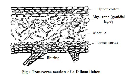
(b) Foliose or Foliaceous lichens (Leafy lichens) : They are leaf like lobed structure which attached to substratum by rhizoid like organs, e.g., Parmelia,, Paltigera.
(c) Fructicose or Filamentous lichens : They are branched shrubby lichens but small base e.g., Cladonia, Usnea.

# (ii) Internal structure : The bulk portion of lichen thallus is formed by fungal partner. The alga constitutes about 5% of the lichen body. Internally the lichens are of two types
(a) Homoiomerous Thalli : Algal cells and fungal hyphae are uniformly dispersed throughout the thallus, e.g., Collema.
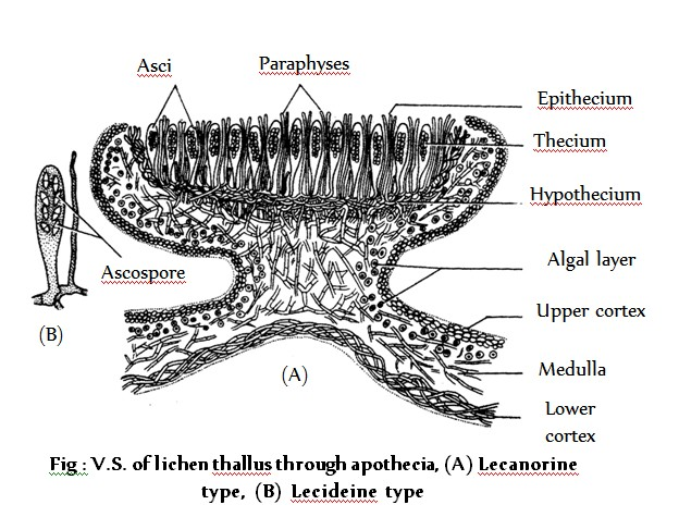
(b) Heteromerous thalli : The algal cells are restricted to algal zone only. In these forms fungal component is dominant. Usually the heteromerous thalli show 4 distinct zones.

- Upper cortex : Formed by compactly interwoven hyphae either without interspaces between them or interspaces filled with gelatinous substances. A cuticle like layer is present on the surface. In some species e.g., Parmelia breathing pores are present.
- Algal layer : Present just below the upper cortex forming photosynthetic zone of the thallus. This layer is also called gonidial layer.
- Medulla : Occurs nearly in the middle of the thallus beneath the algal layer the hyphae are loosely interwoven in this layer.
- Lower cortex : Comprising of closely packed dark coloured hyphae Rhizoids arise from this layer.

Special structures and propagules

The following specialized structures and propagules are associated with lichen thalli :
(i) Breathing pores : The upper surface of some lichens, particularly the foliose lichens is provided with pores. Here the fungal hyphae are loosely arranged. They help in aeration.
(ii) Cyphellae : These are small, almost circular depressions present on the lower side of the thallus. The medullary hypae are not exposed through these depressions due to the presence of corticating hyphae. They are meant for exchange of gases e.g., Sticta. Similar structures without any cortical border are called pseudocyphellae.
(iii) Cephalodia : These are gall like outgrowths present on the upper surface of the thallus. They are distinguishable into cortex and medulla with similar fungal but different algal components from that of the main thallus. Lichens having two algal and one fungal partner are called diphycophilous. The cephaloida are meant for retaining moisture e.g, Peltigera.
(iv) Isidia : These are coral like, simple or branched outgrowths present on the upper surface of the thallus. They have the same algae and fungal component as that of the main thallus. They help in photosynthesis as also in vegetative propogation e.g., Parmelia, Peltigera.
(v) Soredia : It is a powdery mass comprising both algal and fungal components formed in a postule like structure called soralium. The soralia arise from the algal zone lying just below the upper cortex e.g., Physia, Parmelia.

Reproduction

Lichens reproduce both by asexual and sexual methods.

# (i) Asexual reproduction
(a) Fragmentation : The main thallus breaks into small pieces which grow as independent thalli.
(b) Rejuvination : Plants like Cladonia show this unique phenomenon. It becomes young again. The older parts of the thallus die whereas the young branches continue to grow.
(c) Isidia : These are small superficial outgrowths on the surface of lichen thallus. They enclose the same alga as present in the thallus and covered by continuous cortex. Their function is to increase the photosynthesis by increasing the surface area. They get detached from the thallus, disseminate by wind and grow into new thalli.
(d) Soredia : This is a powdery mass formed in a postule like structure called soralium. Each soredium forms a new thallus under favourable conditions.
(e) Conidia : In serveral lichens the fungal component forms conidia on conidiophores. They form new fungal mycelium which with suitable algal component form the lichen.
(f) Pycniospores : The conidia formed in a flask shaped structures lying embedded in the thallus (pycnidia) are called as pycniospores. The pycniospores form new fungal mycelium which consitute the lichen on coming in contact with suitable algal component.

# (ii) Sexual reproduction : Sexual reproduction in lichen is performed only or mainly by its fungal component. So, the structure of the reproductive organs is dependent upon the type of their fungal partner.
The ascolichens reproduce sexually by forming sex organs. The female sex organ is called ascogonium and the male, pycnidium. The ascogonium is a multicelled structure coiled in its basal region. The terminal region is some what eract and called trichogyne. The ascogonium remains embedded in the thallus. The pycnidia acting as male sex organs are called spermogonia. They are pitcher shaped structures that lie embedded in the thallus. The conidia formed in the spermogonia act as spermatia. Some sterile hyphae also emerge out of the ostiole. The spermatia are colourless tiny structures of varying shapes, they are disseminated by wind. Finally, they attach themselves to the sticky tip of the trichogyne. This is followed by plasmogamy and migration of the male nucleus to the female structure. Ascogenous hyphae now develop from the fertilized cell of ascogonium. The asci develop by crozier formation and karyogamy occurs inside the ascus mother cell. This is followed by meiosis and mitosis resulting in the formation of eight ascospores inside the ascus. Simultaneously, the surrounding hyphae also develop and as a result fruiting body called ascocarp or ascomata is formed. The ascocarp may be an apothecium or perithecium.

Accordingly, the ascolichens are divided into two groups namely gymnocarpae and pyrenocarpae respectively. While the apothecia are cup- shaped structures e.g., Physcia, the perithecia are pitcher shaped e.g., Acrocordia. The fruiting body is internally distinguishable into three zones.
(a) Thecium : It is the fertile zone comprising fertile asci and sterile paraphyses.
(b) Epithecium : It is the zone formed by the tips of paraphyses projecting beyond the asci.
(c) Hypothecium : It is the zone formed by loosely packed hyphae lying below the thecium.

An apothecium has two types of margins, proper and thalline. The proper margin is formed by fungal hyphae only whereas the thalline margin includes the algal component also. Accordingly, we differentiate two types of apothecia.
(a) Lecideine type : They have only the proper margin e.g., Lecidea.
(b) Lecanorine type :They have both proper as well as thalline margin e.g., Lecanora.
The sterile tissue lying in between the asci is sometimes called hamathecium. The asci dehisce releasing the ascospores. The ascospores germinate to form the fungal hyphae. On coming in contact with the suitable algal component, they constitute the lichen.

Economic importance

# (i) Pioneer of vegetation : Lichens are considered as pioneers in initiating a plant succession on rocks. These are the first plant group which play an important role in the formation of the soil. So lichens are called as formers of nature or soil builders. Crustose being the first followed by foliose and finally fructicose lichens.

# (ii) Food and Fodder : Reindeer moss (Cladonia rangiferina) of the arctic region is the used as food for reindeer and cattle. Iceland moss (Cetraria icelandica) is ground up and mixed with wheat and made into cakes in Iceland. Rock tripe (Umbillicaria) has been eaten by travellers when they face starvation in actic regions. Evernia is used by Egyptians for making bread and Umbilicaria esculenta is regarded as a delicacy in Japan. Species of Parmelia are used as curry powder in India.

# (iii) Medicinal uses : Dog lichen (Peltigera canina) was used as medicine for hydrophobia in ancient days and Lungwort (Lobaria pulmonoria) was used for the diseases of lungs. Usnic acid obtained from Usnea (old man's beard) and Cladonia sp. is used as broad spectrum antibiotic. It is effective against gram positive bacteria. Lobaria pulmonaria, Cetraria icelandica are used in respirotory diseases particularly T.B., Roccella montagnei in angina, Peltigera canina in hydrophobia, Parmelia sexatilis in eipilepsy and Usnea barbata in urinary troubles.

# (iv) In perfumery : Ramalina and Evernia, having sweet scented thalli, are used in the preparation of Dhup, Havan Samagri and soap. Perfumes are extracted from Evernia prunastri and Lobaria pulmonaria.

# (v) In tanning and dying : Lichens like Cetraria icelandica and Labaria pulmonaria are used in tanning. A red dye is obtained from Ochrolechia sp. whereas Parmelia sp. yield a brown dye. Litmus used as acid-base indicator is obtained from Roccella montagnei and Lasallia pustulata. An orchill dye is obtained from Roccella and Leconara which is purified as orcein and used as a biological stain.

# (vi) In brewing and distilling : The lichens contain carbohydrates in the form of lichenin. Cetraria islandica and Cladonia rangiferina (yield upto 66% of the polysaccharides) are used to obtain alcohol in Sweden and Russia.

# (vii) Indicators of air pollution : Lichens are very sensitive to and grow only in free atmosphere. So lichens like Usnea are used as indicators of air pollution.

# (viii) As poison : Some lichens are poisonous also such as Letharia vulpina due to vulpinic acid, Cetraria juniperina due to pinastrinic acid, Parmelia molliuscula due to selenium, Xanthoria parietina due to beryllium and Everina furfuracea due to chlorine.

# (ix) Other uses : Some lichen yield important chemicals e.g., salazinic acid (Ramalina siliquosa), Lecanoric acid (Parmelia subrudecta) and squamatic acid (Cladonia crispata) etc. In hot season, Usnea gets dry and becomes highly inflamable. It easily catches fire and causes forest fires.

Viruses

The term 'virus' has been derived from Latin, which means poison or venom or viscous fluid. These are highly controversial group of microscopic objects (smaller than bacteria, mycoplasma, nostoc, etc.) and are most perfect obligate intracellular parasites of the world. They remain inactive outside a living host but become active inside the host and multiply in it. They represent a transitional form of life between non–living and living world. Nowadays, these are defined as "Viruses are infectious nucleoproteins". The definition given by Green (1935) states that the viruses are the smallest units showing reproductive properties considered typical of life.
According to Bawden (1949), "Viruses are obligate parasites, too small to be seen."
Luria (1953) defined virus as "Sub-microscopic entities capable of being introduced into specific living cells and reproducing inside such cells only. "Single virus is called 'Virion', most of the plant virus are RNA virus. Must of the animal virus are DNA virus.

Important discovery of virus

(i) Carolous causius (1576) recorded first viral disease in tulips.
(ii) A. Mayer (1886) found a disease in tobacco caused by virus and called it tobacco mosaic disease.
(iii) D. Ivanowski (1892), a Russian Botanist, discovered the infectious nature of the viruses. He was the person, who discovered the virus. He found that juice of an infected tobacco when filtered through bacteria–proof filter, caused disease in healthy plants of tobacco.
(iv) Beijerinck (1898) repeated the same experiment and called it infectious fluid–“contagium vivum fluidum”, therefore, he first used the word Virus.
(v) Popper (1908) reported poliomyelitis virus.
(vi) W. Twort (1915) and D. Herelle (1917) discovered bacteriophages, a kind of virus which infected bacteria and destroyed them.
(vii) M. Schelsinger (1933) for the first time isolated a virus by using the technique of ultracentrifugation
(viii) W. M. Stanley (1935) first time isolated tobacco mosaic virus (TMV) in crystalline form and showed that crystals were made up of proteins. Nobel prize was awarded to him for this work.
(ix) Bawden and Pirie (1938) purified TMV and found it to be a nucleoprotein containing RNA.
(x) Saffermann and Morris (1963) discovered Cyanophages that infect blue–green algae

Nature of viruses

Viruses are regarded as intermediate between non-living entities and living organisms. It is very difficult to ascertain whether they are living or non-living. Some characters of viruses suggest their non-living nature whereas many other characters suggest their living nature. The two views are listed below –
(i) Viruses are non-living : The following characters state that they are non-living.
(a) Viruses have no complete cellular structure. They are not surrounded by cell membrane or cell wall.
(b) They do not show cellular metabolism and lack respiration.
(c) They possess high specific gravity unlike living organisms.
(d) Viruses are active only when they are inside the living host cells. Out side the host, they are good as chemical substances. Thus, they do not have their independent existance.
(e) Stanley (1935) isolated the viruses in a crystalline form and kept for a long period. In this form they neither grow nor reproduce but remain in a crystalline form. This phenomenon has not been observed in any living organism.
(f) The viruses can be precipitated just like chemical substances.
(g) Postulates of Robert Koch are not true for the viruses. Virus cannot grow in “invitro” condition in lab.
(ii) Viruses are living organisms : The following characters state that they are living organisms –
(a) They have definite shape and morphology like that of a living organism.
(b) They possess genetic material (DNA or RNA), which determine their structure and development. Genetic material passes from generation to generation in usual manner.
(c) All viruses are intracellular obligate parasite and attack specific hosts. The bacteriophages recognise the real bacterial surface. The viruses produce characteristic symptoms on their particular host.
(d) They show property of mutation.
(e) They show irritability and respond to environmental conditions such as heat, ultraviolet rays, humidity, drought, alcohol, etc.
(f) They can grow inside the host and multiply enormously showing one of the most important property of living organisms.

Chemical composition

Chemically viruses are nucleoproteins. They are made up of central core of nucleic acid. Nucleic acid is only one, either DNA or RNA. This nucleic acid (DNA or RNA) represents the genetic characters of virus. TMV has RNA (like most plant viruses have) 10% RNA and 90% protein is present in influenza virus and PSTV (Potato Spindle Tuber Viroid) also has RNA but it does not have capsid (protein coat). Plant viruses contain RNA but in cauliflower mosaic virus contain DNA. Bacteriophages contain DNA and almost half animal viruses contain RNA and half contain DNA. But it is called that often animal viruses contain DNA. Cancer causing viruses reovirus contain both RNA and DNA, protein is not genetic material and is left outside the host cell and nucleic acid enters into the cell during the process of infection virus can performs division only inside the host cell. Therefore, their metabolism is not independent. Some animals viruses may be coverd by a lipo-proteinaceous envelope Such viruses are called as Lipovirus. Influenza virus also contains carbohydrates. The envelope is made up of virus protein and host cell lipids. One specific feature of the envelope is that it is covered with projections called spikes. Only some enzymes are detected in viruses such as – Lysozyme in bacteriophages, transcriptase in vaccinia virus, reverse transcriptase and DNA or RNA polymerase in retroviruses.

Shape & Size

Shape : There is variation in shapes of viruses. Viruses are always found in geometrical shapes. The virion may be spherical, oval, rod–like, brick–shaped or tadpole–like in shape. On the basis of shape viruses have been placed in the following categories.
(i) Straight, rigid rods with helical architecture, e.g. TMV, Barley stripe mosaic virus (BSMV).
(ii) Long flexous thread–like rods, e.g. Potato latent mosaic, Wheat streak mosaic virus.
(iii) Polyhedral virions, e.g. Turnip yellow mosaic, Tobacco ring spot virus.
(iv) Tadpole like – Bacteriophages.
(v) Spherical – Influenza virus.
(5) Size : Viruses have a long range of size. They range from 10 micromt to more than 300 micromt in size. The virus of foot and mouth disease (FMD) of animals is smaller than the largest protein molecule. The size of some viruses is as follows–Alfalfa mosaic virus is about 17 micromt. Turnip yellow mosaic virus 20–30 micromt, Maize stunt virus 240-50 micromt and Hydrangia spot virus is 44 micromt * 16 micromt. Smallest virus is foot and mouth disease virus (10 micromt). Largest virus is smallpox virus – variola (250 micromt).

General structure of virus

Structurally viruses are made up of envelope, capsid, nucleoid and occasionally one or two enzymes.
(i) Envelope : Some viruses possess an outer thin loose covering, called envelope. It is composed of proteins (from virus), lipids and carbohydrates (both from host). The smaller subunits of envelope are called peplomers. Envelope is mainly found in some animal viruses (e.g., Herpes Virus, HIV, Influenza virus, Rous sarcoma virus) and rarely in some plant viruses (e.g., Potato yellow dwarf virus) and bacterial viruses (e.g., Pseudomonas Z). The viruses, which do not possess envelope, are called naked.
(ii) Capsid : It is the protein coat that surrounds the central portion of nucleoid and enzymes (if present). The capsid consists of a specific number and arrangement of small sub-units called capsomeres. These sub-units possess antigenic properties.
(iii) Nucleoid : The nucleic acid present in the virus is called nucleoid. It is the infective part of virus which utilizes the metabolic machinery of the host cell for synthesis and assembly of viral components. The genetic material of viruses are of four types :
(a) Double stranded DNA (ds DNA) : Occur in Herpes virus, Pox virus, Cauliflower mosaic virus (linear), Hepatitis–B virus (circular).
(b) Single stranded DNA (ss DNA) : Occur in Coliphage fd (linear), coliphage (cyclic).
(c) Double stranded RNA (ds RNA) : Occur in Reo virus, wound tumour virus.
(d) Single stranded RNA (ss RNA) : Occur in Tobacco mosaic virus, Influenza virus, Foot and Mouth virus, Polio virus, Retroviruses (e.g. HIV), etc.

Tobacco mosaic virus (TMV)

It was discovered by the Russian worker D. Ivanowski. Franklin etal (1957) described the ultrastructure of (TMV) – It is a rod–shaped virus having a central core of RNA surrounded by protein coat (capsid) to form the nucleocapsid. The nucleocapsid may be naked or may be surrounded by a loose membranous envelope. It is composed of a number of subunits called capsomeres. The protein coat (capsid) consists of 2130 identical subunits (capsomeres). The protein is 94% and RNA is only 6%. In the entire length a single RNA molecule runs in the form of spiral coils. The molecular weight of RNA molecule is about 2 million. It provides a code which controls the amino acid sequence in the capsid. This virus measuring 300 × 18 nm.

Bacteriophage

The viruses which attack bacteria are called bacteriophages. In outline they look like tadpole or sperm. The body can be divided into a hexagonal head neck and a tail. The hexagonal head has a central core of DNA, which is surrounded by protein coat. The DNA is double helix, coiled molecule, about 50 m in length. It is different from cellular DNA because it has hydroxymethyl cytosine (HMC) in place of cytosine. The cylindrical tail is hollow and is entirely made up of proteins. At the end of this, there are six long threads called tail fibres or caudal fibres. These fibres help the virus while attaching to bacteria. Bacteriophage contain lysozyme enzyme. The water of holy Ganga river contains bateriophage therefore bacteria cannot grow in the water of Ganga.

Cyanophages

Generally some of the viruses are found which attack on blue green algae. Sofferman and Morris (1963) reported 11 filamentous forms of blue green algae (Lyngbya, plactonema and phormidium, hence called LPP-1) which were attacked by viruses. These viruses are usually called cyanophages. Cyanophages contain DNA as their genetic material. These viruses resemble with bacteriophages in morphology and behaviour. The cyanophages which acttack Nostoc (called N-1) and Anabaena variabilis (called An-1) are tadpole like whereas those which attack oscillatoria are rodshaped.

# Mycophages : Some fungi such as, Mushrooms, Penicillium, etc have also been found to be infected by viruses. These are isometric in shape and contain double stranded RNA.
# Phycophages : These are virus which attack on Algae.

 
SiteLock