Centrosome /Centrosome
(1) Discovery : Centrosome was first discovered by Van Benden (1887) and structure was given by T. Boveri.
(2) Occurrence : It is found in all the animal cell except mature mammalian RBC’s. It is also found in most of protists and motile plant cells like antherozoids of ferns, zoospores of algae and motile algal forms e.g., Chlamydomonas but is absent in prokaryotes, fungi, gymnosperms and angiosperms.
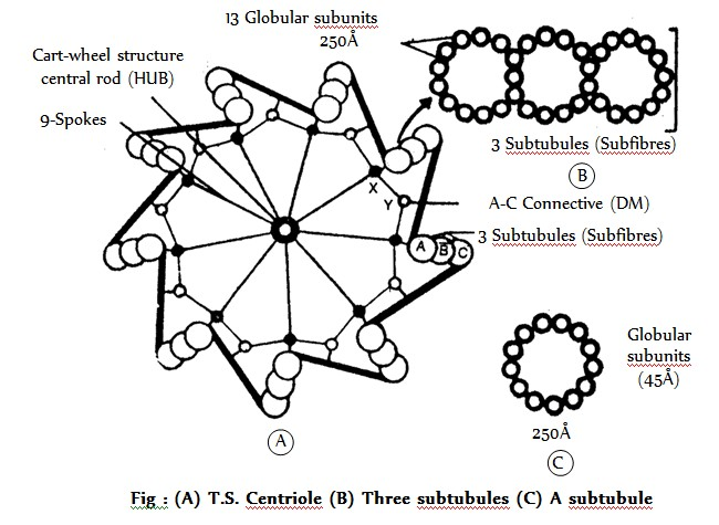
(3) Structure : Centrosome is without unit membrane structure. It is formed of two darkly stained granules called centrioles, which are collectively called diplosome. These centrioles are surrounded by a transparent cytoplasmic area called centrosphere or Kinetoplasm. Centriole and centrosphere are collectively called centrosome. Before the cell division the centrioles at each pole of the spindle. The two centrioles are situated at to each other. Each centriole is a microtubular structure and is formed of microtubules arranged in 9 + 0 manner (all the 9 microtubules are peripheral in position).
Each microtubule is a triplet and is formed of three subtubules which are called A, B and C. A subtubule is about 45Å thick and is formed of 13 parallel protofilaments while each of B and C subtubule is formed of 10 parallel protofilaments. Each protofilament is formed of a row of , -tubulin dimers. C sub-tubule of each microtubule is linked to A sub-tubule of adjacent microtubule by a dense material (DM) strand called A-C linker, so all the microtubules are tilted at . Each microtubule is about 250Å in diameter.
Inside the microtubules, there is an intra-centriolar or cart-wheel structure which is formed of a central hub (about 25Å in diameter) and 9 radial spokes or radial fibres. Each radial spoke ends into a dense material (DM) thickening, called X-body or foot which is further linked to A-subtubule. Between two adjacent X-bodies there is another DM-thickening, called Y-body, which is linked to X-body on either side and to A-C linker on outer side.
Centriole is rich in tubulin and ATPase. Centriole can replicate but has no DNA. Centrioles replicate in phase of interphase of cell cycle but do not initiate cell division.
(4) Chemical composition : Centrosome is lipoproteinaceous structure. The microtubules of centriole are composed of protein tubulin and some lipids. They are rich in ATPase enzyme.
(5) Origin : The daughter centriole is formed from the pre-existing centriole in of interphase so called self-replicating organelle.
(6) Functions
(i) The centrioles help organising the spindle fibres and astral rays during cell division. Therefore, they are called microtubules organising centres. The cells of higher plants lack centrioles and still form a spindle.
(ii) They provide basal bodies which give rise to cilia and flagella.
(iii) The distal centriole of a spermatozoan give rise to the axial filament of the tail.
(2) Occurrence : It is found in all the animal cell except mature mammalian RBC’s. It is also found in most of protists and motile plant cells like antherozoids of ferns, zoospores of algae and motile algal forms e.g., Chlamydomonas but is absent in prokaryotes, fungi, gymnosperms and angiosperms.
(3) Structure : Centrosome is without unit membrane structure. It is formed of two darkly stained granules called centrioles, which are collectively called diplosome. These centrioles are surrounded by a transparent cytoplasmic area called centrosphere or Kinetoplasm. Centriole and centrosphere are collectively called centrosome. Before the cell division the centrioles at each pole of the spindle. The two centrioles are situated at to each other. Each centriole is a microtubular structure and is formed of microtubules arranged in 9 + 0 manner (all the 9 microtubules are peripheral in position).
Each microtubule is a triplet and is formed of three subtubules which are called A, B and C. A subtubule is about 45Å thick and is formed of 13 parallel protofilaments while each of B and C subtubule is formed of 10 parallel protofilaments. Each protofilament is formed of a row of , -tubulin dimers. C sub-tubule of each microtubule is linked to A sub-tubule of adjacent microtubule by a dense material (DM) strand called A-C linker, so all the microtubules are tilted at . Each microtubule is about 250Å in diameter.
Inside the microtubules, there is an intra-centriolar or cart-wheel structure which is formed of a central hub (about 25Å in diameter) and 9 radial spokes or radial fibres. Each radial spoke ends into a dense material (DM) thickening, called X-body or foot which is further linked to A-subtubule. Between two adjacent X-bodies there is another DM-thickening, called Y-body, which is linked to X-body on either side and to A-C linker on outer side.
Centriole is rich in tubulin and ATPase. Centriole can replicate but has no DNA. Centrioles replicate in phase of interphase of cell cycle but do not initiate cell division.
(4) Chemical composition : Centrosome is lipoproteinaceous structure. The microtubules of centriole are composed of protein tubulin and some lipids. They are rich in ATPase enzyme.
(5) Origin : The daughter centriole is formed from the pre-existing centriole in of interphase so called self-replicating organelle.
(6) Functions
(i) The centrioles help organising the spindle fibres and astral rays during cell division. Therefore, they are called microtubules organising centres. The cells of higher plants lack centrioles and still form a spindle.
(ii) They provide basal bodies which give rise to cilia and flagella.
(iii) The distal centriole of a spermatozoan give rise to the axial filament of the tail.