Biology CELL - THE UNIT OF LIFE

Nucleus

(1) Definition : (Karyon = Nucleus) The nucleus also called director of the cell. It is the most important part of the cell which directs and controls all the cellular function.

(2) Discovery : The nucleus was first observed by Robert Brown (1831). Nucleus plays determinative (in heredity) role in cell and organism, that was experimentally demonstrated by Hammerling (1934) by conducting surgical experiments with green marine unicelled algae Acetabularia.

(3) Occurence : A true nucleus with definite nuclear membrane and linear chromosome, is present in all the eukaryotes except mature mammalian RBCs, sieve tube cell of phloem, tracheids and vessels of xylem. The prokaryotes have an incipient nucleus, called nucleoid or prokaryon or genophore or false nucleus or bacterial chromosome.

(4) Number : Usually there is a single nucleus per cell i.e. mononucleate condition, e.g. Acetabularia.
(i) Anucleate (without nucleus) : RBCs of mammals, phloem sieve tube, trachids and vessels of xylam.
(ii) Binucleate : e.g. Ciliate, Protozoans like Paramoecium.
(iii) Polynucleate : e.g. fungal hyphae of Rhizopus, Vaucheria. Polynucleate condition may be because of fusion of a number of cells. i.e. syncytium, coconut endosperm or by free nuclear divisions without cytokinesis i.e. coenocyte.

(5) Shape : It varies widely, generally spherical e.g. cuboidal germ cells, oval e.g. columnar cells of intestine, bean shaped in paramoecium, horse-shoe shaped in vorticella, bilobed, e.g. WBCs (acidophils), 3 lobed e.g. basophil, multilobed e.g. neutrophils, long and beaded form (moniliform) e.g. stentor and branched in silk spinning cells of platy phalyx insect larva.

(6) Size : The size of nucleus is variable i.e. 5 – 30. In metabolically active cells size of the nucleus is larger than metabolically inactive cells. The size depends upon metabolic activity of the cells. It is directly proportional to number of chromosomes.

(7) Chemical composition of nucleus
Proteins = 80% (65% acidic, neutral and enzymatic proteins; 15% basic proteins-histones)
DNA = 12%
RNA = 5%
Lipids = 3%
Enzymes like polymerases are abundantly present and help in synthesis of DNA and RNA. Minerals like and are present in traces.

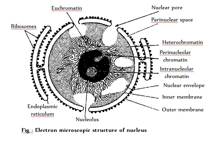
(8) Ultrastructure : The nucleus is composed of following structure
(i) The nuclear membrane
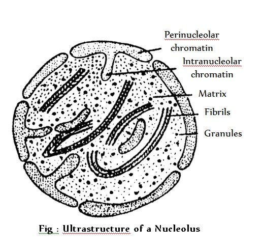
(ii) The nucleous.
(iii) The nuclear sap or nucleoplasm.
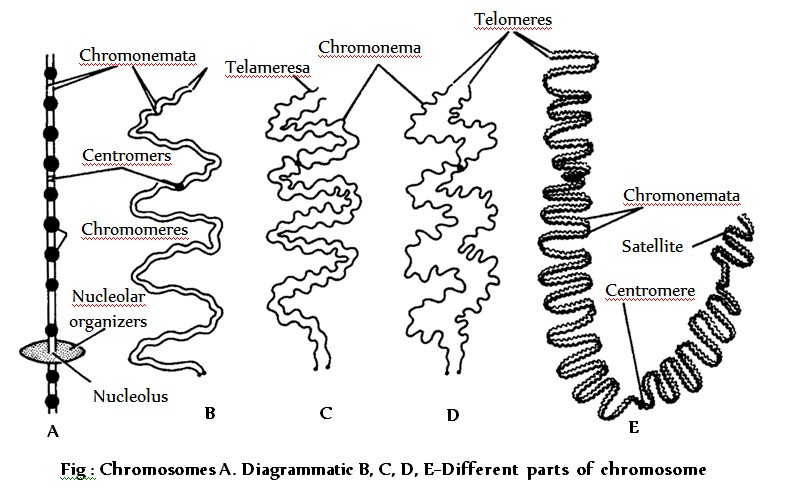
(iv) The chromatin fibres.

The Nuclear Membrane or Karyotheca

(i) Definition : It is defined as a regulatory envelope which controls the nucleo-cytoplasmic interacitons and exchange of materials.

(ii) Discovery : Nuclear membrane, also called nuclear envelope or nucleolemma or karyotheca, was first discovered by Erclab (1845).

(iii) Structure : It is a bilayered envelope. Each membrane is about 90 Å thick lipoproteinous and trilaminar. Outer membrane, called ectokaryotheca, is studded with ribosomes on its cytoplasmic surface and is continuos with RER at some points. Inner membrane, called endokaryotheca, is without ribosomes and is internally lined by electron-dense material of protein fibres called fibrous or nuclear lamina nuclear cortex or hoeny comb layer (about 300 Å thick). Two membranes are separated by a fluid-filled intermembranous perinuclear space (about 100-300Å). Nuclear membrane contains following structure.
(a) Nuclear pore : Nuclear membrane is porous and has 1,000-10,000 octagonal nuclear pores. Each nuclear pore is about 400-1,000 Å in diameter (average size is 800 Å). The number and size of the nuclear pores depend upon the needs of the cell. Nuclear pores are interspaced at about 1000-1500 Å. Each nuclear pore is fitted with a cylindrical structure, called annulus (with a lumen of 500 Å) and both collectively form the pore complex or pore basket. Annulus has 8 micro-cylinders (each about 200 Å in diameter and with a lumen of 50 Å) in its wall. It also encloses a channel having nucleoplasmin for the movement of substances. Annulus acts as a diphragm and regulates the size of the nuclear pore.
(b) Nuclear blebbing : The nuclear envelope shows evagination. As a result, blebs are formed which are pinched off. This phenomenon is called blebbing. The nuclear vesicles so formed are thought to give rise to mitochondria, plastids, etc. Blebbing may also occur from the outer unit membrane only. A row of these blebs move towards the periphery. As a result of deposition of matrix material in between these blebs, and annulate lamella is formed. The annulate lamellae is thought to give rise to ER cisternae.

(iv) Origin : It is formed by the fusion of ER elements during the telophase of cell division.

(v) Functions
(a) It regulates the nucleo-cytoplasmic interactions.
(b) It allows the passage of inorganic ions and small organic molecules.
(c) It helps in pinocytosis and phagocytosis of large sized molecules .
(d) It allows passage of ribosomal subunits, RNAs and proteins through nuclear pores.
(e) It maintains the shape of the nucleus.
(f) Fibrous lamina strengthens the nuclear envelope. It also helps in dissolution and reformation of nuclear membrane during cell division.

The nucleolus (Little nucleus)

(i) Discovery : Nucleolus was first observed by Fontana (1781) in the skin cells of an eel. Term ‘nucleous’ was coined by Bowman (1840). Its light microscopic structure was given by Wagner (1840).

(ii) Position : It is generally associated with nucleolar organizer region (NOR) of the nucleolar chromosomes. It is absent in muscle fibres, RBC, yeast, sperm and prokaryotes.

(iii) Number : Generally, a diploid cell is with two nucleoli but there are five nucleoli in somatic cell of man and about 1000 nucleoli in the oocytes of Xenopus.

(iv) Structure : (De Robertis et. al 1971). A nucleolus is distinguishable into following regions :-
(a) Chromatin : The nucleolus is surrounded by perinucleolar chromatin. Heterochromatic intrunsions are also seen in the nucleolus which constitutes the intranucleolar chromatin.
(b) Pars fibrosa : Fibrils of 80 – 100 Å size form a part of the nucleolus.
(c) Pars granulosa : Granules of 150 – 200 Å diameter constitute the granular part of the nucleolus. They appear like vesicle with a light central core. The granules may be joined by filament forming a beaded primary nucleolonema. The fibrils may also be associated to it. The primary nucleolonema may further coil to form the secondary nucleolonema.
(d) Pars amorpha : The granules and the fibrils lie dispered in an amorphous proteinaceous matrix. Nucleolus contains large amount of proteins mainly phosphoproteins. There are no histones proteins. RNA methylase, an enzyme that transfers methyl groups to the RNA bases has been localized in nucleolus. Nucleolus is stained by “pyronine”. It is not bounded by any limiting membrane. Fibrillar region of nucleolus is called secondary constriction or nucleolar organising region (NOR) and this region directs the synthesis of rRNA. Ribosomes are assembled here as such it is also called ribosome producing machine or factory. Ribosomal units so formed are joined together by thin filament (rRNA) forming a structure like string of beads and it is called “nucleonema”.

(v) Chemical composition : Nucleolus is mainly formed of RNA and non histone acidic proteins. It is a storehouse of RNA.

(vi) Origin : A nucleolus is formed at specific sites, called the nucleolar organizers, present on certain chromosomes region (NOR).

(vii) Functions
(a) It is seat of biogenesis of rRNA and also stores rRNA.
(b) It plays important role in spindle formation during cell division.
(c) It receives the ribosomal proteins from the cytoplasm, combines the rRNAs and ribosomal proteins to form ribosomal subunits.
Nucleoplasm : It is also called karyolymph. It is transparent, homogenous, semifluid, colloidal, ground substance present inside the nuclear membrane in which nuclear chromatin and nucleoli are embedded. Chemically it contains. Nucleoplasm is also known as protoplasm of nucleus.

(i) Nucleic acid : Monomer nucleotides of DNA and RNA
(ii) Proteins : Basic proteins (nuclear protamines and nucleohistones and acidic proteins (non-histone)
(iii) Enzymes : DNA polymerase, RNA polymerase, NAD synthetase, nucleoside triphosphatase, and pyruvic acid kinase, etc.
(iv) Minerals : Phosphorus, potassium, sodium, calcium, magnesium, etc.
(v) Ribonucleoproteins : Contain perichromatin granules and interchromatin granules. Histone proteins are basic because they contain arginine in much amount e.g. .
The nucleoplasm helps in maintaining the shape of nucleus formation of spindle protein of NAD, ATP, DNA, RNAs and ribosomal subunits. Plasmosome and karyosome combindly called “amphinucleoli”.
Chromatin fibres /Nuclear chromatin : The nucleoplasm contains many thread like, coiled and much elongated structures which take readily the basic stains such as “basic fuschin”. These thread like structures are known as chromatin fibre. They are uniformly distributed in the nucleoplasm. They are observed only in the “interphase stage”. Chromatin fibres are made of chromosomes. In resting nondividing eukaryotic cells the genome is nucleoprotein complex and it is called chromatin.

Chromatin

(1) Definition : During interphase, chromatin threads are present in the form of a network called chromatin reticulum. At the time of cell division, these thread like structures of chromatin become visible as independent structures, called chromosomes.

(2) Structure of chromosome : Each chromosome consists of two coiled filaments throughout its length called chromonemata by Vejdovsky. These have bead like structures called chromomeres which bear genes. Chromatid is a half chromosome or daughter chromosome. The two chromatids are connected at the centromere or primary constriction. Primary constriction (centromere) and secondary constriction gives rise to satellite. The secondary constriction consists of genes which code for ribosomal RNA and nucleolus hence it is called as “nucleolar organizer region”. Chromosomes having satellite are called SAT chromosomes.
The ends of chromosomes are called “telomeres” (which do not unite with any other structure). A tertiary constriction is also present in chromosomes, which perhaps helps in recognition of chromosomes.
In 1928 Emile Heitz developed a technique for stainning of chromosomes. Chromosomes can be stainned with acetocarmine or fuelgen (basic fuschin) there are two types of regions are seen :–

# (i) Heterochromatin : It is formed of thick regions which are more darkly stained than others areas. It is with condensed DNA which is transcriptionally inactive and late replicating. It generally lies near the nuclear lamina. Heterochromatin are of two types : –
(a) Facultative heterochromatin : Temporarily inactivated chromatin and forms 2.5% of the genome.
(b) Constitutive hetrochromatin : Permanently inactivated chromatin and generally ground near centromeres.

# (ii) Euchromatin : It is true chromatin and is formed of thin, less darkly stained areas. It is with loose DNA which is transcriptionally active and early replicating.

(3) Chemical chomposition : DNA - 40%. Histone – 50%. Other (acid) Proteins – 8.5%. RNA – 1.5%. Traces of lipids, Ca, Mg and Fe. Histone are low molecular weight basic proteins which occur alongwith DNA in ratio. Nonhistone chromosomal or NHC proteins are of three types– structural, enzymatic and regulatory. Structural NHC proteins form the core or axis of the chromosome. They are also called scaffold proteins. Enzymatic proteins form enzymes for chemical transformation, e.g., phosphates, RNA polymerase, DNA polymerase. Regulatory proteins control gene expression. HMG (high mobility group) proteins get linked to histones for releasing DNA to express itself.

 
SiteLock