The study of blood vessels is called Angiology. The blood vessels are of following types :
(i) Arteries (ii) Capillaries (iii) Veins
Vasa vasorum : Supply blood to the wall of large blood vessels.
# (i) Arteries : Thick walled, carrying oxygenated blood (deoxygenated in pulmonary artery) from heart to various parts of body. These blood vessels are grouped as Aorta which branches to form arteries which further divides into thinner branches called arterioles inside the organ. Average diameter of arteriole is 120 m. the arterioles further divide into smaller vessels called meta-arterioles (70 m) which divide into capillaries. At the beginning of capillary, the arterioles posses circular muscles called precapillary sphincter which regulates flow of blood into the capillaries which is called vasomotion.
- Muscleless end of meta-arteriole is called thoroughfare channel or preferential channel.
- The largest artery is dorsal / abdominal aorta (systemic aorta).
- Elastic or conducting arteries receive blood from heart and do not provide it to any organ rather they provide blood to other atreries and are pressure reservoirs of blood.
- Muscular arteries show vasoconstriction and vasodilation and provide blood to the organs.
- Anastomosis : If more than one arteries are supplying to one organ then branches of these arteries unite to form a network called - - Anastomosis. It provides many collateral or alternate pathways of blood supply. So, if there is blocking of any artery, it will not lead to necrosis.
- End arteries : In organs like heart, branches of different arteries do not unite rather they terminate due to which the alternate pathways are not available. In such cases, blocking of any artery leads to necrosis of related part of organ. To develop alternate pathway in such conditions is called as By pass surgery.
# (ii) Capillaries : Smallest blood vessels, discovered by Marcello Malpighl (also layered nucleated squamous epithelial cells called endothelium resting on a basement membrane. Diameter of capillary is about 8. These are also called as exchange vessels as they are the site of exchange of material between blood and tissue because of least barrier in them. The capillaries can be grouped into two categories :
(a) Arteriolar capillary : Which supplies nutrition, respiratory gases etc. to the body cells.
(b) Veinular capillaries : Which collect the metabolic wastes from the body cells.
Capillaries possess abour 5% of total body blood and are present near almost all cells of body in the intercellular spaces. The tissues which are devoid of intercellular spaces are also devoid of capillary. They are called avascular tissues.
- Capillaries are surrounded by cells of connective tissue called pericapillary cells. Some of these cells are contractile and phagocytic in nature and are called Rouget cells or pericytes.
- Continuous capillaries are without fenestra/aperture, hence are less permeable. These are present in organs such as lungs, muscles, connective tissues and brain tissues.
- Fenestrated capillaries possess apertures/fenestra and are found in those organs where there is maximum need of permeability such as endocrine glands, intestinal villi, cavities of brain, kidney, ciliary body of eye.
- Sinusoids are irregularly dilated capillaries found in organs where there is decrease in flow rate such as liver, spleen, bone marrow, parathyroid, pituitary gland. In liver, sinusoids are branches of venules and open into venules while in other organs, they originate from arteriole and unite to form venules.
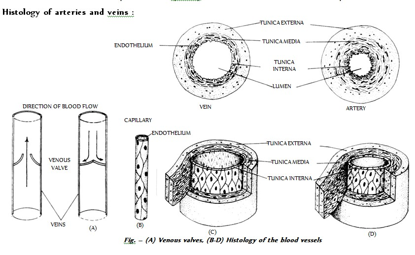
# (iii) Veins : These are thin walled, carrying deoxygenated blood (oxygenated in pulmonary vein) from tissues to the heart. Venules, smallest branches, unite to form veins which in turn unite to form vena cava. The largest vein is inferior vena cava/post caval. Varicose veins is stout, blood filled painful veins specially of the limbs due to defective watch pocket valves.
The study of blood vessels is called Angiology. The blood vessels are of following types :
(i) Arteries (ii) Capillaries (iii) Veins
Vasa vasorum : Supply blood to the wall of large blood vessels.
# (i) Arteries : Thick walled, carrying oxygenated blood (deoxygenated in pulmonary artery) from heart to various parts of body. These blood vessels are grouped as Aorta which branches to form arteries which further divides into thinner branches called arterioles inside the organ. Average diameter of arteriole is 120 m. the arterioles further divide into smaller vessels called meta-arterioles (70 m) which divide into capillaries. At the beginning of capillary, the arterioles posses circular muscles called precapillary sphincter which regulates flow of blood into the capillaries which is called vasomotion.
- Muscleless end of meta-arteriole is called thoroughfare channel or preferential channel.
- The largest artery is dorsal / abdominal aorta (systemic aorta).
- Elastic or conducting arteries receive blood from heart and do not provide it to any organ rather they provide blood to other atreries and are pressure reservoirs of blood.
- Muscular arteries show vasoconstriction and vasodilation and provide blood to the organs.
- Anastomosis : If more than one arteries are supplying to one organ then branches of these arteries unite to form a network called - - Anastomosis. It provides many collateral or alternate pathways of blood supply. So, if there is blocking of any artery, it will not lead to necrosis.
- End arteries : In organs like heart, branches of different arteries do not unite rather they terminate due to which the alternate pathways are not available. In such cases, blocking of any artery leads to necrosis of related part of organ. To develop alternate pathway in such conditions is called as By pass surgery.
# (ii) Capillaries : Smallest blood vessels, discovered by Marcello Malpighl (also layered nucleated squamous epithelial cells called endothelium resting on a basement membrane. Diameter of capillary is about 8. These are also called as exchange vessels as they are the site of exchange of material between blood and tissue because of least barrier in them. The capillaries can be grouped into two categories :
(a) Arteriolar capillary : Which supplies nutrition, respiratory gases etc. to the body cells.
(b) Veinular capillaries : Which collect the metabolic wastes from the body cells.
Capillaries possess abour 5% of total body blood and are present near almost all cells of body in the intercellular spaces. The tissues which are devoid of intercellular spaces are also devoid of capillary. They are called avascular tissues.
- Capillaries are surrounded by cells of connective tissue called pericapillary cells. Some of these cells are contractile and phagocytic in nature and are called Rouget cells or pericytes.
- Continuous capillaries are without fenestra/aperture, hence are less permeable. These are present in organs such as lungs, muscles, connective tissues and brain tissues.
- Fenestrated capillaries possess apertures/fenestra and are found in those organs where there is maximum need of permeability such as endocrine glands, intestinal villi, cavities of brain, kidney, ciliary body of eye.
- Sinusoids are irregularly dilated capillaries found in organs where there is decrease in flow rate such as liver, spleen, bone marrow, parathyroid, pituitary gland. In liver, sinusoids are branches of venules and open into venules while in other organs, they originate from arteriole and unite to form venules.
# (iii) Veins : These are thin walled, carrying deoxygenated blood (oxygenated in pulmonary vein) from tissues to the heart. Venules, smallest branches, unite to form veins which in turn unite to form vena cava. The largest vein is inferior vena cava/post caval. Varicose veins is stout, blood filled painful veins specially of the limbs due to defective watch pocket valves.