Biology BODY FLUIDS AND CIRCULATION

Human Circulatory System - Structure of Human Heart And Blood Vessels

This system is concerned with the circulation of body fluids to distribute various substances to various body parts.

Functions of Circulatory System :
 Transport of various substances such as nutrients, waste products, respiratory gases, metabolic intermediates (Such as lactic acid from muscle to liver), vitamins hormones etc.
 Regulation of body pH by means of buffer, body temperature homeostasis, water balance etc.
 Prevention of disease by means of antibodies and antitoxins.
 Support or turgidity to certain organs like penis and nipples.

Types of Circulation

Circulatory system in various groups of animals can be classified as follows :
(i) Intracellular circulation : Occurs inside the individual cells where the distribution of substances is through cyclosis of cell cytoplasm. Example – Protozoans.
(ii) Extracellular circulation : When the distribution of the substances occurs inside the body through extracellular or intracellular fluids. This is of following types –
(a) Extra organismic circulation : When the water of the external environment circulate through body. This is also called as water circulation system. Example – canal system in porifera, water vascular system in Echinoderms and gastrovascular system in coelenterates.
(b) Intra-organismic circulation : It involves circulation of body fluids. It is of following types :
(1) Parenchymal circulation : In platyhelminthes, the fluid filled spaces present in the mesodermal parenchyma tissue between body wall and internal organs are used in the distribution of substances.
(2) Coelomic circulation : Coelomic fluid is concerned with the transport of substances. Example – pseudocoelomic fluid in the roundworms and haemolymph in Arthropods.
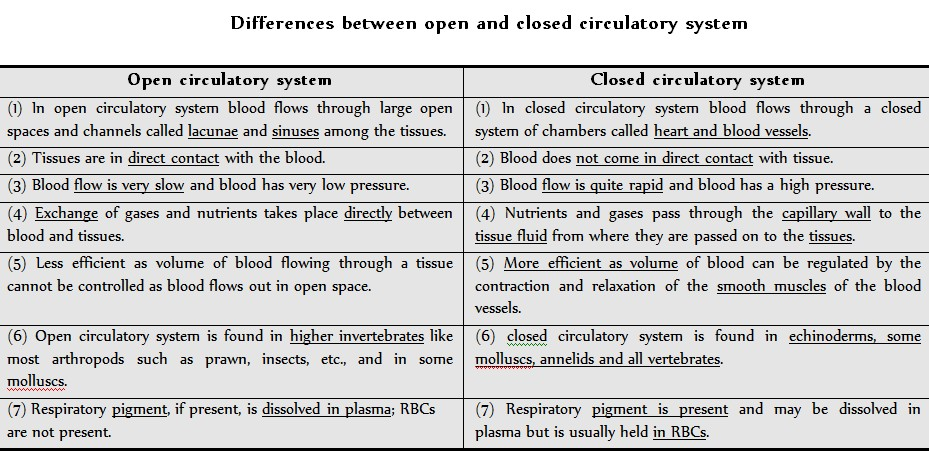
(3) Blood vascular system : It contains blood and a pumping structure (heart) for circulation of materials inside the body. It is of following types –
(i) Open circulatory system
(ii) Closed circulatory system

Circulatory system in multicellular animals

(i) In protozoans : Distribution of nutrients takes place by cyclosis (streaming movement) of cytoplasm.
(ii) In poriferans : The vascular system of poriferans is the canal system. A simplest canal system involves ostia (mouth), spongocoel and on osculum (Anus).

# Route followed by water current in sponges given in attached fig.

(iii) In coelenterates : Hydra has a single large internal cavity called coelenteron or gastrovascular cavity. It has single opening the mouth. It also extends into the hollow tentacles. It lacks a mesodermal epithelial covering (peritoneum) and a coelomic fluid. It is concerned with first extracellular and then intracellular digestion of food.

(iv) In platyhelminthes : Vascular system is absent but circulation occurs with the help of parenchyma hence called parenchymal circulation. Example – Fasciola hepatica.

Heart

(i) Shape and position : Reddish, roughly conical, highly muscular, mesodermal hollow organ of the size of one’s first. Its average weight in males is about 300 gm. and in females about 250 gm. It lies behind the sternum in the mediastinum space of thoracic cavity in between the two lungs. The broader base faces upward and backward. The narrower apex is directed downward, forward and slightly towards left, lying between 5th and 6th ribs and rests on the diaphragm.

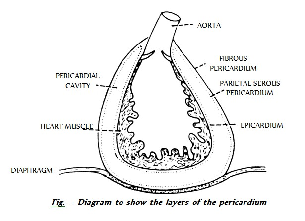
(ii) Protective covering : Heart is enclosed in a tough, 2 layered fibroserous sac, the pericardium. The outer layer is non-distensible fibrous pericardium and inner layer is thin serous pericardium which further consists of outer parietal layer (attached to fibrous pericardium) and inner visceral layer (adhered to the heart).
Between the parietal and visceral layers, occurs a narrow potential space, the pericardial cavity which is derived from coelom and is filled with serous pericardial fluid for frictionless movement and protection from shock and mechanical injury.

Histology :

The heart wall consists of connective tissue, blood vessels and cardiac muscle fibres in 3 different layers ¬– Epicardium, Myocardium and Endocardium.
(a) Endocardium : Innermost layer lining the cavity of heart and consisting of endothelium of squamous cells resting on thin basement membrane of loose connective tissue.
(b) Myocardium : Middle, highly vascular layer, composed of cardiac muscle fibres joined together by intercalated disc. The connective tissue in myocardium acts as cardiac skeleton. Endocardium is thickest where the myocarduim is thinnest and vice-versa.
(c) Epicardium : Visceral pericardium, joined to myocardium by connective tissue.

External structure

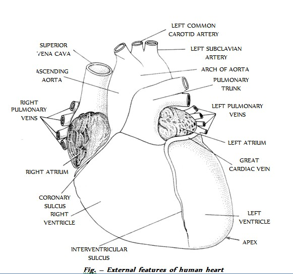
Human heart is 4-chambered and is divided by septa into two halves – right and left. Each half has one darker, thin walled auricle in the broader upper region and one lighter, thick-walled ventricle in the narrower lower region.

# (a) Auricles (Atria) : Two in no., demarcated externally from ventricles by irregular groove called coronary sulcus and from each other by interatrial sulcus. When atria contract, small curtain like flaps called auricular appendages or appendices project from sides of auricles and overhang the corresponding ventricles.

# (b) Ventricles : Two in no. demarcated externally from each other by an oblique groove called interventricular sulcus which contains coronary blood vessels. The right ventricle does not reach apex.

# (c) Sinus venosus and conus arteriosus : Sinus venosus and conus/truncus/bulbus arteriosus are accessory chambers in the heart of lower vertebrates (fishes and amphibians). In rabbit, sinus venosus is formed in the embryo but later it becomes a part of wall of right auricle.

- In frog, sinus venosus spreads upon most of the dorsal side of heart and conus arteriosus lies obliquely upon the ventral surface of right atrium.

Internal structure

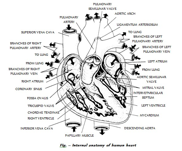
(a) Auricles : Atria are thin walled. They act as reserviors for blood entering the heart. Right auricle is bigger than left auricle and both are separated by a myomembranous partition called Interatrial or interauricular septum. During embryonic stage, at the place of this septum, there are present septum primum and septum secondum having a gap (aperture) called foramen ovalis between them. From the opening of inferior vena cava upto foramen ovalis, there is a flap called Eustachian flap which prevents the blood in the foetal heart go to lungs because in foetal life, lungs are not functional purification of blood is done by placenta.
At the time of birth, there is closure of foramen ovalis but there remains depression on posterior part of the right surface of interauricular septum in rabbit. In man this depression is present on both the side. because of least regenerative power in human being. The depression towards right atrium is called fossa ovalis and depression towards left atrium is called fossa lunata.

# PFO (Patient Foramen Ovalis) or septal defect : In case there is no closure of foramen ovalis, then disease is called PFO. In this condition, there is mixing of blood after birth which gives bluish appearance to the body called as Cyanosis. Such child is called Blue Baby.
The inner surface of auricles is smooth. A network of muscular ridges called musculi pectinati or trabeculi pectinati occurs internally in the region of the auricular appendages and give comb like appearance.

(b) Ventricles : The right and left ventricles are demarcated by an interventricular septum which is obliquely curved towards right, so that the left ventricle is larger than right one. However, the cavity of left ventricle is relatively smaller and nearly circular because the myocardium of left ventricleis 3 times thicker than right ventricle whose cavity is larger and somewhat crescentic.
The walls of the ventricles are internally raised into a number of thick, muscular, column shaped projections called columnae carnae or trabecular carnae; and a few large muscular elevations called papillary muscles or musculli papillares which are 3 in right ventricle and 2 in left ventricle. These muscles act as anchors for chordae tendinae.

# Chordae tendinae : Numerous, strong, inelastic thread like tendons present in the mammalian heart but absent in frog. One end of these threads is attached to the cusps of A.V. valves and the other end to the papillary muscles of the ventricles. These muscles contract during ventricular systole and pull the valves downwards, thus, preventing their everting into atria. The chordae tendinae hold the valves in place.

# Regurgitation : If there is weeakening of papillary muscles or breaking of chordae tendinae, then AV valves revert into auricles. So, blood goes in opposite direction, it is called regurgitation. Sometimes, there is narrowing of valves. So, there remains gap between the valves which causes regurgitation.

#Moderator band : Right ventricle contains a prominent muscular trabeculum called moderator band which extends from the interventricular septum to anterior papillary muscle.

Major blood vessels associated with heart

The blood vessels that enter or leave the heart are called Great Blood Vessels.
# (a) Superior vena cava or precaval : Brings deoxygenated blood from head and upper parts of the body into the right auricle through an opening which is single in human and cat and two in rabbit as there are 2 precavals – right and left in rabbit. In frog, right and left precavals open into sinus venosus.

# (b) Inferior vena cava or post caval : Drains deoxygenated blood from middle and lower parts of the body into the right auricle through a single opening which is bordered by a membranous, falciform fold which is a remnant of the foetal valve of Eustachian. In frog, post caval opens into sinus venosus.

# (c) Coronary sinus : Returns deoxygenated blood from heart wall into right auricle through a single opening.

# (d) Pulmonary vein : Four pulmonary veins, two from each lung, carry oxygenated blood from the lungs and open into the left auricle through four openings. In rabbit, the pulmonary veins open in the left auricle through 2 openings.

# (e) Pulmonary aorta/arch : Arises from upper left corner of right ventricle through a single opening and divides into right and left pulmonary arteries which carry deoxygenated blood to the lungs for oxygenation.

# (f) Systemic aorta : Arises from upper right corner of left ventricle through a single opening and has 3 regions – ascending aorta, arch of aorta and descending aorta. It distributes oxygenated blood to various body parts except lungs.
Ligamentum arteriosus : During foetal life, because the lungs are non-functional hence blood of pulmonary aorta comes into systemic aorta through a small duct called ductus botalli or ductus arteriosus soon after birth, deposition of elastin fibre blocks this duct, forming a new structure called ligamentum botalli or ligamentum arteriosus.

- PDA (Patient Ductus Arteriosus) : If the ligamentum arteriosus remains open, the condition is called PDA. In this case, there is mixing of blood which leads to blue baby.

# Valves : The valves present in the mammalian heart are tendinous cords.
- (a) Eustachian valve : Present on the opening of inferior vena cava (post caval) in the right auricle in rabbit, whereas in human, the vestige of eustachian valve is present over the opening of post caval vein. It allows the passage of blood in right auricle.
- (b) Haversian valve : Present in human but absent in rabbit. It is present over the opening of precaval vein and allows the passage of blood in right auricle.
- (c) Thebesian or coronary valve : Present over the opening of coronary sinus in right auricle in mammals and allows the passage of blood in right auricle.
- (d) Atrio-ventricular valves : Auricles open into the respective ventricles through wide passages called auriculo ventricular apertures or A.V. apertures which are guarded by one-way A.V. valves or parachute valves and are located dorsally or posteriorly. There are 2 types of valves in mammals.
(1) Right A.V. valve or Tricuspid valve : Present between right auricle and right ventricle. It consists of 3 membranous flaps or cusps.
(2) Left A.V. valve or Bicuspid or Mitral valve : Present between left auricle and left ventricle. It consists of 2 flaps or cusps. The bicuspid
valve resembles mitre or topi of bishop, hence, also called as Mitral valve.

# The upper edges of the flaps are attached to the margins of the A.V. apertures while the lower edges project freely into the ventricles. The free edges of these flaps are connected by chordae tendinae to he papillary muscles of the ventricles. These valves allow the passage of blood from auricles into ventricles but prevent backflow.
In frog, the A.V. valves are semilunar type and not of cuspid type. There is single row of A.V. valves due to single ventricle.

- (e) Semilunar valves : At the base of pulmonary arch and systemic aorta, three membranous, pocket-shaped flaps called semilunar valves are present which are set in a ring with their cavities directed away from the ventricles. They allow the passage of blood from ventricles to respective blood vessels, but prevent the return of blood.

Physiology of heart

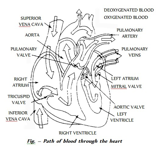
The heart pumps blood to all parts of the body. The deoxygenated blood is drained into right auricle through superior and inferior venae cavae and coronary sinus whereas the pulmonary veins carry oxygenated blood from lungs to the left auricle. This is called as Auricular circulation. About 70% of the auricular blood passes into the ventricles during diastole. This phase is called diastasis. The rest of 30% of blood passes into the ventricles due to auricular systole (contraction). In this way, blood reaches the ventricles and is called ventricular filling. During ventricular systole (which starts first in left ventricle than in right ventricle), the pressure increases in the ventricles, thus, forcing the oxygenated blood from left ventricle into systemic aorta and deoxygenated blood from right ventricle into pulmonary aorta. The systemic arch distributes the oxygenated blood to all the body parts except lungs while pulmonary aorta carries the deoxygenated blood to lungs for oxygenation.

# During foetal life, heart receives and pumps mixed blood and hence, it can be compared with transitional heart, the eustachian flap, in the foetus directs the blood of right atrium towards left atrium through foramen ovalis. From left atrium, blood reaches left ventricle from which the systemic aorta arise. An iliac artery arise from this aorta from the internal branches of illiac artery, two umbilical arteries arise which come out of body through naval and reach placenta where exchange of gases takes place. A single umbilical vein arises from placenta and enters the foetal body through naval and reaches the liver to give some blood to it and some blood to inferior vena cava. Inferior vena cava already possess impure blood. So, there is mixing of blood. In foetus, pure blood is there only in umbilical vein (allantoic vein). Umbilical cord is a tube possessing a jelly like connective tissue (Wharton jelly) along with two umbilical arteries and one umbilical vein.

- (a) Heart beat : The spontaneous and rhythmic contraction and relaxation of the heart to pump out and receive blood to and from the body is called Heart beat. Depending upon the nature of control of the heart beat, hearts are of 2 types –
Neurogenic and Myogenic or autorhythmic.

- (b) Origin and conduction of heart beat : Initiation of heart beat is under special bundles of cardiac muscles called nodal tissue. The cardiac muscles have less actin and myosin. So, structurally they become more a nerve than muscle and functionally they are similar to neurons.

Morphology of nodal tissue

The nodal tissue consists of the following –

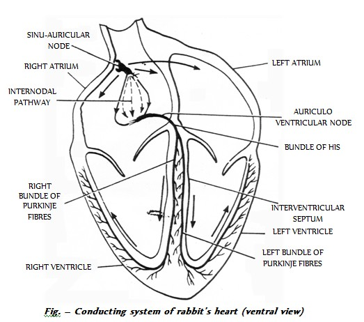
# (i) Sinu-auricular or S.A. node : Also called as pacemaker, node of keith and flack, heart of heart, brain of heart, pulsation centre. It is located in the right wall of right atrium below the opening of superior vena cava. This is the place where sinus venosus is incorporated in the wall of right atrium in the embryo. S.A. node is the main tissue of heart and has highest degree of autrohythmicity (generates beating impulse at the rate of 70-80 times/minute) but least conductivity. The rhythmic impulses produced are called as Sinus rhythmia. In frog S.A. node is present in sinus venosus.

# (ii) Atrio-ventricular node or A.V. node : Also called reserve pacemaker, node of Twara and Aschoff. Discovered by Lewis Kent. It lies in the right atrium near the junction of interauricular and interventricular septum close to the opening of coronary sinus. It is concerned with the conduction of cardiac impulses generated by S.A. node, but it can also generate the impulse at the rate of 40-60/minute. These impulses produced are rhythmic and called nodal rhythmia. In frog, A.V. node is absent.

# (iii) Bundle of His or A.V. bundle : Discovered by His. It arises from A.V. node, descends in the interventricular septum and bifurcates into two branches innervating the wall of right and left ventricle respectively. The myocardium of atria and ventricles are discontinuous and this bundle is the only muscular connection between the two. It is concerned with the conduction of impulse from atria to the tip of ventricle but can also generate impulse at the rate of 35-40/minute. The impulses produced are non-rhythmic.

# (iv) Purkinje fibres : Numerous, modified muscle fibres which act as sympathetic nerve fibres. They arise from branches of bundle of His and provide impulse to myocardium of ventricles. They can also generate non-rhythmic impulse at a rate of 30-35/minute.

Working of nodal tissue

S.A. node spontaneously initiates a wave of contraction which is conducted along the tracts of special muscle fibres called internal pathways over both the auricles at a rate of 1m/sec. The impulse generated travels first in the right atrium than in left atrium. So, right atrium contracts first but the contraction ends simultaneously in both atria. As the musculatures of atria and ventricles are discontinuous and are separated by a septum of fibrous connective tissue, called annular pad in mammals, the wave of contraction is received by A.V. node from myocardium of atria and is provided to bundle of His. The impulses reach the A.V. node about 0.03 seconds after their origin from S.A. node. The A.V. node generates a fresh wave of contraction which passes over both the ventricles along the bundle of His and its ramifications at the rate of 1.5 to 4 m/sec. The Purkinje fibres bring about the contraction of ventricles from the apex of heart which passes quickly towards the origin of pulmonary and systemic arches forcing blood into them.

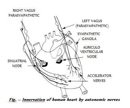
S.A. node not only acts as pacemaker but also establishes the basic rhythm at which the heart beats. In case of degeneration of S.A. node, A.V. node can generate impulse but it will lead to abnormal beating (arrhythmia). The failure of atrial impulse to pass into ventricles for a few seconds to few hours is called ventricular escape or stokes-adams syndrome leading to delayed pick up of heart beat. In such conditions, artificial pacemaker (Lithium Battery) is placed underneath the patient’s chest.
Ectopic pacemaker : If any cardiac muscle other than the conducting tissue (nodes) generates impulse, then extra beats are heard. Such muscles are called Ectopic pacemaker.

In mammals, conducting system of the heart has S.A. node, A.V node and complicated system of conducting fibres. But in frog, it has only S.A. node and system of conducting fibres is simple.
Heart beat rate : Heart beat/minute or number of cardiac cycles/minute. Example – frog-64/min., rabbit-200/min., human-70-80/min. Females have higher heart rate than males.

Normal heart beat rate - Rhythmia
Abnormal heart rate - Arrhythmia
Decrease in heart rate - Bradycardia
Increase in heart rate - Tachycardia

Blood vessels

The study of blood vessels is called Angiology. The blood vessels are of following types :
(i) Arteries (ii) Capillaries (iii) Veins
Vasa vasorum : Supply blood to the wall of large blood vessels.

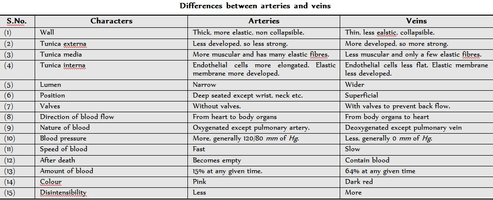
# (i) Arteries : Thick walled, carrying oxygenated blood (deoxygenated in pulmonary artery) from heart to various parts of body. These blood vessels are grouped as Aorta which branches to form arteries which further divides into thinner branches called arterioles inside the organ. Average diameter of arteriole is 120 m. the arterioles further divide into smaller vessels called meta-arterioles (70 m) which divide into capillaries. At the beginning of capillary, the arterioles posses circular muscles called precapillary sphincter which regulates flow of blood into the capillaries which is called vasomotion.
- Muscleless end of meta-arteriole is called thoroughfare channel or preferential channel.
- The largest artery is dorsal / abdominal aorta (systemic aorta).
- Elastic or conducting arteries receive blood from heart and do not provide it to any organ rather they provide blood to other atreries and are pressure reservoirs of blood.
- Muscular arteries show vasoconstriction and vasodilation and provide blood to the organs.
- Anastomosis : If more than one arteries are supplying to one organ then branches of these arteries unite to form a network called - - Anastomosis. It provides many collateral or alternate pathways of blood supply. So, if there is blocking of any artery, it will not lead to necrosis.
- End arteries : In organs like heart, branches of different arteries do not unite rather they terminate due to which the alternate pathways are not available. In such cases, blocking of any artery leads to necrosis of related part of organ. To develop alternate pathway in such conditions is called as By pass surgery.

# (ii) Capillaries : Smallest blood vessels, discovered by Marcello Malpighl (also layered nucleated squamous epithelial cells called endothelium resting on a basement membrane. Diameter of capillary is about 8. These are also called as exchange vessels as they are the site of exchange of material between blood and tissue because of least barrier in them. The capillaries can be grouped into two categories :
(a) Arteriolar capillary : Which supplies nutrition, respiratory gases etc. to the body cells.
(b) Veinular capillaries : Which collect the metabolic wastes from the body cells.
Capillaries possess abour 5% of total body blood and are present near almost all cells of body in the intercellular spaces. The tissues which are devoid of intercellular spaces are also devoid of capillary. They are called avascular tissues.
- Capillaries are surrounded by cells of connective tissue called pericapillary cells. Some of these cells are contractile and phagocytic in nature and are called Rouget cells or pericytes.
- Continuous capillaries are without fenestra/aperture, hence are less permeable. These are present in organs such as lungs, muscles, connective tissues and brain tissues.
- Fenestrated capillaries possess apertures/fenestra and are found in those organs where there is maximum need of permeability such as endocrine glands, intestinal villi, cavities of brain, kidney, ciliary body of eye.
- Sinusoids are irregularly dilated capillaries found in organs where there is decrease in flow rate such as liver, spleen, bone marrow, parathyroid, pituitary gland. In liver, sinusoids are branches of venules and open into venules while in other organs, they originate from arteriole and unite to form venules.

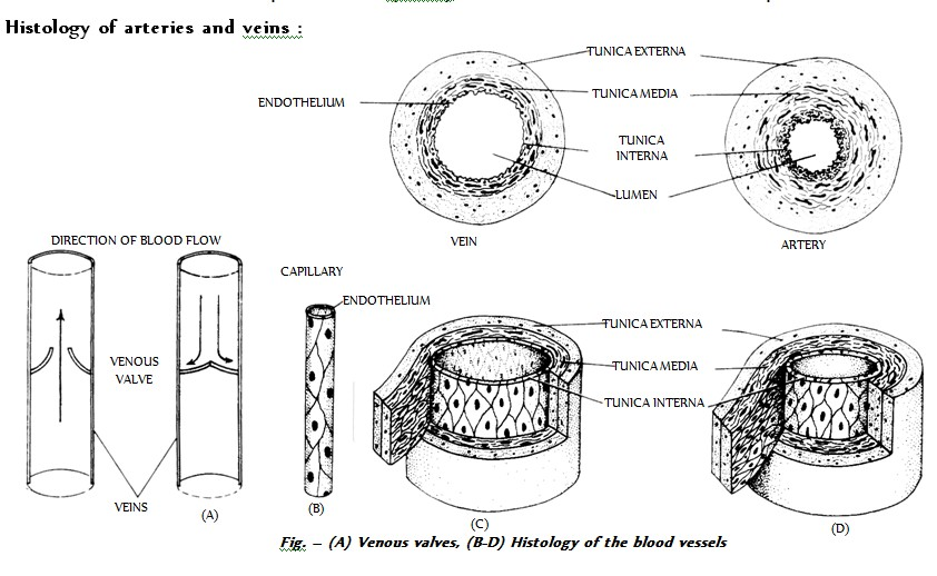
# (iii) Veins : These are thin walled, carrying deoxygenated blood (oxygenated in pulmonary vein) from tissues to the heart. Venules, smallest branches, unite to form veins which in turn unite to form vena cava. The largest vein is inferior vena cava/post caval. Varicose veins is stout, blood filled painful veins specially of the limbs due to defective watch pocket valves.
Histology of arteries and veins

(1) Tunica externa or tunica adventitia : Outermost, fibrous, made up of collagen rich connective tissue and less elastin fibres. The collagen fibres give strength to the blood vessels and prevent their overdilation.
(2) Tunica media : Middle, thickest, made up of smooth involuntary muscle fibres and elastin fibres. This layer is very much variable because number of elastin fibres and muscle fibres depend upon the position of blood vessels from the heart.
(3) Tunica interna or tunica intima : Innermost, thinnest, made up of inner, single layer of simple squamous epithelial cells called endothelium resting on a basement membrane and outer layer of elastic (yellow fibrous) connective tissue. The hollow space in the blood vessel is called lumen.

Blood pressure

The pressure exerted by the blood on the wall of the blood vessels in which it is present is called blood pressure. It is usually measured in brachial artery by an instrument called sphygmomanometer (invented by Riva-Rocci). Arterial blood pressure is of 2 types :

# (1) Systolic blood pressure : It is the pressure exerted by blood on the walls of the blood vessels due to the systole of ventricles and is equal to 120 mm Hg. During ventricular systole, there is expansion in the artery due to the uncoiling of elastic layer. Hence, the pressure is maximum in arteries but gradually decreases in capillaries and veins.

# (2) Diastolic blood pressure : It is the pressure exerted on walls of blood vessels when the ventricles are relaxed. During ventricular diastole, the uncoiled elastic layer recoils leading to normalization of artery. Hence, blood pressure drops down to 80 mm Hg. Thus, blood pressure in normal person is systolic/diastolic pressure i.e. 120/80 mm Hg.

# (3) Pulse pressure : The difference between systolic and diastolic pressures is called pulse pressure and its normal value is 120 – 80 mm Hg = 40 mm Hg. It provides information about the condition of arteries.

# (4) Mean arterial pressure : It is the average pressure of systolic and diastolic pressures. As the blood remains in the systolic phase for shorter period and in the diastolic phase for longer period, the mean pressure of blood lies near the diastolic pressure.
This value varies at different levels of circulation being maximum (100 mm Hg) in the aorta and minimum (0 mm Hg) in the venae cavae under normal conditions.

# Pulse : It is the pressure wave of distension and recoiling felt in the radial artery due to the contraction of left ventricle which force about 70-90 ml of blood in each cardiac cycle to aorta. This perssure wave of contraction travels down to the wall to the arteries and is called the pulse.
- The pulse is measured in the radial artery in the wrist but can be felt in the temporal artery over the temporal bone or the dorsal pedis artery at the bind of ankle. The pulse normally travels at the rate of 5-8 m/second.
Since each heart beat generates one pulse in the arteries so the pulse rate per minute indicates the rate of heart beat. So the normal pulse rate in a normal adult person is 72/minute.
- The normal ratio of systolic pressure to diastolic pressure to pulse pressure is about 3 : 2 : 1.

 
SiteLock