Lymphatic system in human
# (a) Lymph capillaries : Small, thin, lined by endothelium resting on a basement membrane and fine whose one end is blind and other end unites to form lymphatic ducts. These are present almost throughout the body but are absent in brain, eyeball, spinal cord, internal ear, bone marrow etc. Lymph capillaries in the region of small intestine in villi are called “lacteals” which collect chyle which is milky white in colour due to absorbed fat. Lacteals help in the absorption of digested fat.
# (b)Lymphatic ducts or vessels : Numerous, present in various parts of body. These vessels are like veins as they have all the three layers – tunica externa, tunica media and tunica interna, and are provided with watch pocket or semilunar valves but valves are more in number than veins.
# (c) Flow of lymph in lymphatics : Pulsations of lymph hearts in frog create sufficient force to maintain a steady flow of lymph in the lymphatic system. In mammals, the credit for maintaining onwards flow of lymph goes to (i) the “squeezing force” created by the muscles of body wall and internal organs, (ii) the breathing movements of diaphragm and thoracic cage, (iii) mild peristalsis created by smooth muscles of the wall, of lymphatics themselves, and (iv) the pressure created by increasing amount of lymph in the lymphatics. Certain compounds like fats increase the rate of lymph flow and are called lymphata gogue. Blocking of lymph flow causes oedema.
# (d) Types of lymphatic ducts : Two main types :
- (1) Right lymphatic duct : It is the smallest lymphatic duct with the length of approximately 1.25 cm. Its one end is blind and other one opens into right subclavian vein at the junction of right internal jugular vein. It collects lymph from one-fourth of the body (right part of head, neck, thoracic cavity and right arm).
- (2) Left lymphatic duct/thoracic duct : It is the longest lymphatic duct with the length of approximately 38-45 cm. It originates from cisterna chyli and empties into left subclavian vein. It collects lymph from three-fourth part of the body i.e. complete posterior part through cisterna chyli, left part of head, neck, thoracic cavity and left arms.
- (3) Cisterna chyli receptaculum chyli : It is a dilated sac like structure present below the diaphragm in lumbar region at the level of second lumbar vertebra. It collects lymph from posterior part of body i.e. abdomen, pelvic region and hind limbs and drains it in the left lymphatic duct.
It shows inflation and deflation due to the movement of diaphragm which is a passive movement. Hence, it is also called as passive lymphatic artery. It is also called as second heart.
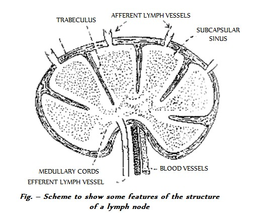
- (4) Lymph nodes or lymph glands : These are the masses of lymphatic tissue and connective tissue (reticular tissue) and are located on the capillaries either solitary or in cluster. Where they are present solitary and in few number, such tissues are called diffused lymphatic tissues and where they are in clusters, they are called tonsils.
# Lymph nodes are covered by capsule of white collagen tissue. Outer region of lymph node is called cortex and inner region is called medulla. In medulla, there are medullary cords, (cord like arrangement of lymphocytes). Cortex possess follicles (clusters of lymphocytes), outer part of which possess T-cells and macrophages while the inner part possess B-cells.
# Lymphadenitis : During infection, central part of follicle shows rapid division and formation of plasma cells. hence, this part is also called reaction centre. The inflammation of lymph nodes in such condition is called Lymphadenitis.
- Some of the common lymph nodes are – Axillary nodes (in armpits), genital (Inguinal) nodes (in pubic region), cervical nodes (in neck region), intercostal nodes (in chest region), lumbar nodes (in lumbar region), iliac nodes (in pelvic region) and payer’s patches (in small intestine). Besides these lymphatic nodes, a number of them are also present near major blood vessels (arteries), specially dorsal aorta.
- Tonsils : Clusters of lymph nodes. They are very often called as policemen. Various tonsils are – Normal tonsils (in pharynx), adenoid tonsils (in nasopharynx), abdominal tonsils (in vermiform appendix) and policeman of intestine (in lamina propria of ileum). Adenoid tonsils are present upto 7 years of age, then they are degenerated. Their swelling is called adenoid. Inflammation of tonsils is called Tonsilitis.
- Haemal lymph node : In many animals some lymph nodes are found to possess red colour, due to the presence of blood in them. In man they are found in the retroperitoneal tissues and also in the mediastinum. In these nodes some of the so-called lymphatic channels contain blood, while the rest of the nodes possesses the same structure as the typical lymph node. Spleen may be regarded as the modified haemal lymph (haemolymph) node. Lymph nodes are located at intervals along its course.
# Function of lymph nodes :
(i) They produce and supply lymphocytes to the blood and as a supportive function the trabeculae carry blood vessels which supply the node.
(ii) They make screening of the lymph by means of phagocytic activity.
(iii) They serve a great defensive role against bacterial infections.
(iv) They temporarily stop the spread of cancer cells as those cells have to penetrate through the lymph vessels to the lymph nodes from where they spread in the body.
(v) They act as mechanical filters to resist the entrance of poisonous substances into circulation.
(vi) They carry out immunological responses. They help in elaboration of antibodies and in the development of immunity.
(vii) Lymph nodes produce gamma-globulin.
# (b)Lymphatic ducts or vessels : Numerous, present in various parts of body. These vessels are like veins as they have all the three layers – tunica externa, tunica media and tunica interna, and are provided with watch pocket or semilunar valves but valves are more in number than veins.
# (c) Flow of lymph in lymphatics : Pulsations of lymph hearts in frog create sufficient force to maintain a steady flow of lymph in the lymphatic system. In mammals, the credit for maintaining onwards flow of lymph goes to (i) the “squeezing force” created by the muscles of body wall and internal organs, (ii) the breathing movements of diaphragm and thoracic cage, (iii) mild peristalsis created by smooth muscles of the wall, of lymphatics themselves, and (iv) the pressure created by increasing amount of lymph in the lymphatics. Certain compounds like fats increase the rate of lymph flow and are called lymphata gogue. Blocking of lymph flow causes oedema.
# (d) Types of lymphatic ducts : Two main types :
- (1) Right lymphatic duct : It is the smallest lymphatic duct with the length of approximately 1.25 cm. Its one end is blind and other one opens into right subclavian vein at the junction of right internal jugular vein. It collects lymph from one-fourth of the body (right part of head, neck, thoracic cavity and right arm).
- (2) Left lymphatic duct/thoracic duct : It is the longest lymphatic duct with the length of approximately 38-45 cm. It originates from cisterna chyli and empties into left subclavian vein. It collects lymph from three-fourth part of the body i.e. complete posterior part through cisterna chyli, left part of head, neck, thoracic cavity and left arms.
- (3) Cisterna chyli receptaculum chyli : It is a dilated sac like structure present below the diaphragm in lumbar region at the level of second lumbar vertebra. It collects lymph from posterior part of body i.e. abdomen, pelvic region and hind limbs and drains it in the left lymphatic duct.
It shows inflation and deflation due to the movement of diaphragm which is a passive movement. Hence, it is also called as passive lymphatic artery. It is also called as second heart.
- (4) Lymph nodes or lymph glands : These are the masses of lymphatic tissue and connective tissue (reticular tissue) and are located on the capillaries either solitary or in cluster. Where they are present solitary and in few number, such tissues are called diffused lymphatic tissues and where they are in clusters, they are called tonsils.
# Lymph nodes are covered by capsule of white collagen tissue. Outer region of lymph node is called cortex and inner region is called medulla. In medulla, there are medullary cords, (cord like arrangement of lymphocytes). Cortex possess follicles (clusters of lymphocytes), outer part of which possess T-cells and macrophages while the inner part possess B-cells.
# Lymphadenitis : During infection, central part of follicle shows rapid division and formation of plasma cells. hence, this part is also called reaction centre. The inflammation of lymph nodes in such condition is called Lymphadenitis.
- Some of the common lymph nodes are – Axillary nodes (in armpits), genital (Inguinal) nodes (in pubic region), cervical nodes (in neck region), intercostal nodes (in chest region), lumbar nodes (in lumbar region), iliac nodes (in pelvic region) and payer’s patches (in small intestine). Besides these lymphatic nodes, a number of them are also present near major blood vessels (arteries), specially dorsal aorta.
- Tonsils : Clusters of lymph nodes. They are very often called as policemen. Various tonsils are – Normal tonsils (in pharynx), adenoid tonsils (in nasopharynx), abdominal tonsils (in vermiform appendix) and policeman of intestine (in lamina propria of ileum). Adenoid tonsils are present upto 7 years of age, then they are degenerated. Their swelling is called adenoid. Inflammation of tonsils is called Tonsilitis.
- Haemal lymph node : In many animals some lymph nodes are found to possess red colour, due to the presence of blood in them. In man they are found in the retroperitoneal tissues and also in the mediastinum. In these nodes some of the so-called lymphatic channels contain blood, while the rest of the nodes possesses the same structure as the typical lymph node. Spleen may be regarded as the modified haemal lymph (haemolymph) node. Lymph nodes are located at intervals along its course.
# Function of lymph nodes :
(i) They produce and supply lymphocytes to the blood and as a supportive function the trabeculae carry blood vessels which supply the node.
(ii) They make screening of the lymph by means of phagocytic activity.
(iii) They serve a great defensive role against bacterial infections.
(iv) They temporarily stop the spread of cancer cells as those cells have to penetrate through the lymph vessels to the lymph nodes from where they spread in the body.
(v) They act as mechanical filters to resist the entrance of poisonous substances into circulation.
(vi) They carry out immunological responses. They help in elaboration of antibodies and in the development of immunity.
(vii) Lymph nodes produce gamma-globulin.