Central nervous system
Central nervous system is made up of brain and spinal cord. CNS is covered by 3 meninges and its wall has two type of matter.
Types of matter : CNS of vertebrates is formed of two types of matter –
- (a) Grey matter : It is formed of cell-bodies and non-medullated nerve fibres.
- (b) White matter : It is formed of only medullated nerve fibres which appear white due to presence of medullary sheath.
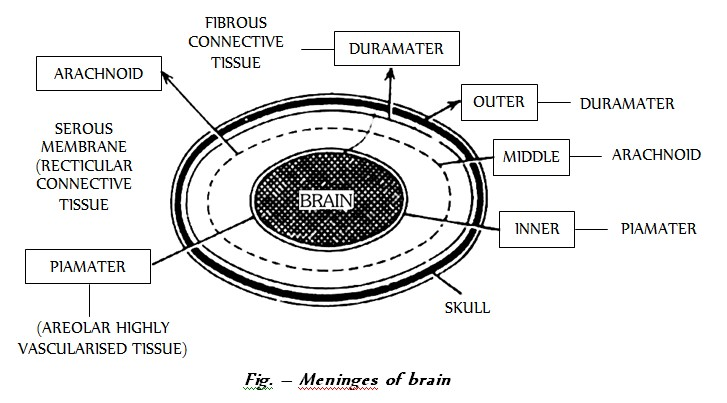
Meninges : The meninges are connective tissue membranes which surround the brain and spinal cord of CNS. In the fishes, there is only one meninx called meninx primitiva. In amphibians, reptiles and birds, the brain is covered by two meninges or membranes : inner pia-arachnoid and outer dura mater. In mammals, CNS is covered by three meninges or membranes :
# (a) Duramater (Dura = tough; mater = mother): Outermost, thick, fibrous, 2-layered meninge. The outer layer adheres to skull at many places while the inner layer follows the major convolutions (sulci and gyri) of the brain and spinal cord. Meningeal artery traverses via duramater. The two layers of duramater are widely separated at some places to form the large sinuses called venous sinus. This drains deoxygenated (= venous) blood from the brain to the large veins that return it to the heard. The space between duramater and the next meninge in succession is called sub-dural space is filled with cerebrospinal fluid and has arachnoid villi in the region of dural space. Similarly the space between the skull and durameter is called epidural space. Duramater extends in the form of straight sulcus between cerebrum and cerebellum posteriorly. Here it is called tentorium.
# (b) Arachnoid (= spider-like web) : It is closely related to duramater on its outside and with piameter on the inside. The space between the arachnoid and piameter is called sub-arachnoid space and is filled with cerebro-spinal fluid.
# (c) Piameter (Pia = soft = tender) : This is the innermost meninge and follows the convolutions of the outer surface of brain and spinal cord. It is highly vascular and penetrates deeply in certain places bringing it with its vasculature and placing it in contact with the ventricles of the brain and neurocoel of spinal cord.
Cerebrospinal fluid : All the ventricles of the brain are continuous and lined by a columnar, ciliated epithelium, the ependyma. They contain lymph-like extracellular fluid called the cerebrospinal fluid (C.S.F.). This fluid is secreted by the choroid plexuses by filtration of blood. The choroid plexuses consist of loose connective tissue of pia mater covered internally by a simple cuboidal epithelim of secretory (glandular) nature. The cerebrospinal fluid slowly flows toward the fourth ventricle by secretion pressure and passes into the spinal cord. Some fluid escapes into the subarachnoid spaces through three pores in the roof of the fourth ventricle in the medulla. From the subarachnoid spaces, the cerebrospinal fluid is transferred to the blood of the venous sinuses. Nervous tissue is without lymphatic vessels.
The cerebro-spinal fluid (CSF) provides
(a) Protection to brain from mechanical socks.
(b) Optimum physiological fluid environment for neural functions e.g. conduction of nerve impulses, transport of aminoacids, sugars, O2 etc.
(c) ‘Relief’ mechanism for the increase in intracranial pressure that occurs with each arterial pulse of blood to brain.
(d) ‘Sink’ like facility for metabolites of brain.
(e) The blood CSF barrier for selective transport process between blood and CSF.
# Major site of CSF formation is choroid plexus, and mid ventricular wall and sub-arachnoid wall also contribute. CSF is cell free, slightly alkaline, and is isotonic to plasma. Rate of formation of C.S.F is 80 ml/hour approx, 1/2 litre per day. Total amount present in and around CNS is 150 ml it means there is atleast 3 times renewal of C.S.F. every day.
Blood brain barrier facilitate maintenance of stable internal environment. Its acts as physiological and pathological barrier as well. Hydrocephalus : The enlargement of head, a pathological condition characterized by an abnormal accumulation of cerebrospinal fluid resulting headache, vomiting, pain and stiffness of the neck.
Increased cerebrospinal fluid may result Meningites.
Meningites may appear due to infection and inflamation of meninges or injury of meninges.
Infection may be viral, bacterial or both. The most common cause of meningitis in the infection of streptococcus and neumoniae, neisseria meningitidis and haemophilus influenzae.
Lumber puncture is done for drainage of excess of cerebrospinal fluid during meningitis.
Cerebro-spinal fluid is formed by choroid plexus (ACP and PCP).
There are three choroid plexus in humans
(a) Lateral choroid plexus : It is in the roof of I and II ventricle.
(b) Anterior choroid plexus : It is in the roof of III ventricle (diacoel).
(c) Posterior choroid plexus or pelochoroida : It is in the roof of IV ventricle.
Oxygen and glucose requirements : Brain controls the functions of our body organs and also provides the qualities of mind – learning, reasoning, and memory. For these activities, brain needs a large and constant energy supply. At any given time, the activities of the brain account for 20% of the body’s consumption of oxygen and 15% of its consumption of blood glucose. Brain deprived of oxygen for just 5 minutes is permanently damaged. Mental confusion results if brain is deprived of glucose.
Types of matter : CNS of vertebrates is formed of two types of matter –
- (a) Grey matter : It is formed of cell-bodies and non-medullated nerve fibres.
- (b) White matter : It is formed of only medullated nerve fibres which appear white due to presence of medullary sheath.
Meninges : The meninges are connective tissue membranes which surround the brain and spinal cord of CNS. In the fishes, there is only one meninx called meninx primitiva. In amphibians, reptiles and birds, the brain is covered by two meninges or membranes : inner pia-arachnoid and outer dura mater. In mammals, CNS is covered by three meninges or membranes :
# (a) Duramater (Dura = tough; mater = mother): Outermost, thick, fibrous, 2-layered meninge. The outer layer adheres to skull at many places while the inner layer follows the major convolutions (sulci and gyri) of the brain and spinal cord. Meningeal artery traverses via duramater. The two layers of duramater are widely separated at some places to form the large sinuses called venous sinus. This drains deoxygenated (= venous) blood from the brain to the large veins that return it to the heard. The space between duramater and the next meninge in succession is called sub-dural space is filled with cerebrospinal fluid and has arachnoid villi in the region of dural space. Similarly the space between the skull and durameter is called epidural space. Duramater extends in the form of straight sulcus between cerebrum and cerebellum posteriorly. Here it is called tentorium.
# (b) Arachnoid (= spider-like web) : It is closely related to duramater on its outside and with piameter on the inside. The space between the arachnoid and piameter is called sub-arachnoid space and is filled with cerebro-spinal fluid.
# (c) Piameter (Pia = soft = tender) : This is the innermost meninge and follows the convolutions of the outer surface of brain and spinal cord. It is highly vascular and penetrates deeply in certain places bringing it with its vasculature and placing it in contact with the ventricles of the brain and neurocoel of spinal cord.
Cerebrospinal fluid : All the ventricles of the brain are continuous and lined by a columnar, ciliated epithelium, the ependyma. They contain lymph-like extracellular fluid called the cerebrospinal fluid (C.S.F.). This fluid is secreted by the choroid plexuses by filtration of blood. The choroid plexuses consist of loose connective tissue of pia mater covered internally by a simple cuboidal epithelim of secretory (glandular) nature. The cerebrospinal fluid slowly flows toward the fourth ventricle by secretion pressure and passes into the spinal cord. Some fluid escapes into the subarachnoid spaces through three pores in the roof of the fourth ventricle in the medulla. From the subarachnoid spaces, the cerebrospinal fluid is transferred to the blood of the venous sinuses. Nervous tissue is without lymphatic vessels.
The cerebro-spinal fluid (CSF) provides
(a) Protection to brain from mechanical socks.
(b) Optimum physiological fluid environment for neural functions e.g. conduction of nerve impulses, transport of aminoacids, sugars, O2 etc.
(c) ‘Relief’ mechanism for the increase in intracranial pressure that occurs with each arterial pulse of blood to brain.
(d) ‘Sink’ like facility for metabolites of brain.
(e) The blood CSF barrier for selective transport process between blood and CSF.
# Major site of CSF formation is choroid plexus, and mid ventricular wall and sub-arachnoid wall also contribute. CSF is cell free, slightly alkaline, and is isotonic to plasma. Rate of formation of C.S.F is 80 ml/hour approx, 1/2 litre per day. Total amount present in and around CNS is 150 ml it means there is atleast 3 times renewal of C.S.F. every day.
Blood brain barrier facilitate maintenance of stable internal environment. Its acts as physiological and pathological barrier as well. Hydrocephalus : The enlargement of head, a pathological condition characterized by an abnormal accumulation of cerebrospinal fluid resulting headache, vomiting, pain and stiffness of the neck.
Increased cerebrospinal fluid may result Meningites.
Meningites may appear due to infection and inflamation of meninges or injury of meninges.
Infection may be viral, bacterial or both. The most common cause of meningitis in the infection of streptococcus and neumoniae, neisseria meningitidis and haemophilus influenzae.
Lumber puncture is done for drainage of excess of cerebrospinal fluid during meningitis.
Cerebro-spinal fluid is formed by choroid plexus (ACP and PCP).
There are three choroid plexus in humans
(a) Lateral choroid plexus : It is in the roof of I and II ventricle.
(b) Anterior choroid plexus : It is in the roof of III ventricle (diacoel).
(c) Posterior choroid plexus or pelochoroida : It is in the roof of IV ventricle.
Oxygen and glucose requirements : Brain controls the functions of our body organs and also provides the qualities of mind – learning, reasoning, and memory. For these activities, brain needs a large and constant energy supply. At any given time, the activities of the brain account for 20% of the body’s consumption of oxygen and 15% of its consumption of blood glucose. Brain deprived of oxygen for just 5 minutes is permanently damaged. Mental confusion results if brain is deprived of glucose.