Restriction Enzymes
In the year 1963, the two enzymes responsible for restricting the growth of bacteriophage in Escherichia coli were isolated. One of these added methyl groups to DNA, while the other cut DNA. The later was called restriction endonuclease.
The first restriction endonuclease–Hind II, whose functioning depended on a specific DNA nucleotide sequence was isolated and
characterised five years later. It was found that Hind II always cut DNA molecules at a particular point by recognising a specific sequence of six base pairs. This specific base sequence is known as the recognition sequence for Hind II. Besides Hind II, today we know more
than 900 restriction enzymes that have been isolated from over 230 strains of bacteria each of which recognise different recognition sequences.
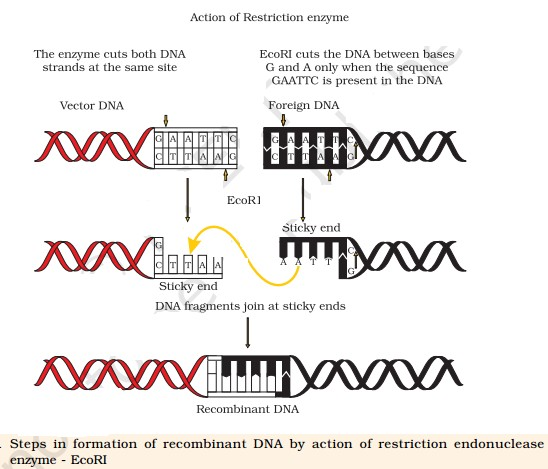
The convention for naming these enzymes is the first letter of the name comes from the genus and the second two letters come from the species of the prokaryotic cell from which they were isolated, e.g., EcoRI comes from Escherichia coli RY 13. In EcoRI, the letter ‘R’ is derived from the name of strain. Roman numbers following the names indicate the order in which the enzymes were isolated from that strain of bacteria.
Restriction enzymes belong to a larger class of enzymes called nucleases. These are of two kinds; exonucleases and endonucleases.
Exonucleases remove nucleotides from the ends of the DNA whereas, endonucleases make cuts at specific positions within the DNA.
You may have realised that normally, unless one cuts the vector and the source DNA with the same restriction enzyme, the recombinant vector molecule cannot be created.
The first restriction endonuclease–Hind II, whose functioning depended on a specific DNA nucleotide sequence was isolated and
characterised five years later. It was found that Hind II always cut DNA molecules at a particular point by recognising a specific sequence of six base pairs. This specific base sequence is known as the recognition sequence for Hind II. Besides Hind II, today we know more
than 900 restriction enzymes that have been isolated from over 230 strains of bacteria each of which recognise different recognition sequences.
The convention for naming these enzymes is the first letter of the name comes from the genus and the second two letters come from the species of the prokaryotic cell from which they were isolated, e.g., EcoRI comes from Escherichia coli RY 13. In EcoRI, the letter ‘R’ is derived from the name of strain. Roman numbers following the names indicate the order in which the enzymes were isolated from that strain of bacteria.
Restriction enzymes belong to a larger class of enzymes called nucleases. These are of two kinds; exonucleases and endonucleases.
Exonucleases remove nucleotides from the ends of the DNA whereas, endonucleases make cuts at specific positions within the DNA.
You may have realised that normally, unless one cuts the vector and the source DNA with the same restriction enzyme, the recombinant vector molecule cannot be created.