Muscle is a specialised tissue of mesodermal origin. About 40-50 per cent of the body weight of a human adult is contributed by muscles.
They have special properties like excitability, contractility, extensibility and elasticity. Muscles have been classified using different criteria, namely location, appearance and nature of regulation of their activities. Based on their location, three types of muscles are identified :
(i) Skeletal
(ii) Visceral
(iii) Cardiac.
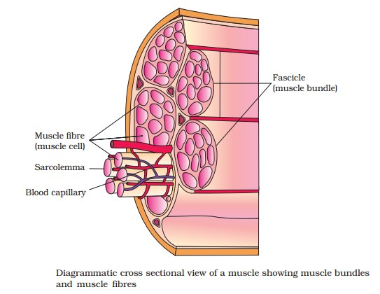
# Skeletal muscles are closely associated with the skeletal components of the body. They have a striped appearance under the microscope and hence are called striated muscles. As their activities are under the voluntary control of the nervous system, they are known as voluntary muscles too. They are primarily involved in locomotory actions and changes of body postures.
# Visceral muscles are located in the inner walls of hollow visceral organs of the body like the alimentary canal, reproductive tract, etc. They do not exhibit any striation and are smooth in appearance. Hence, they are called smooth muscles (nonstriated muscle). Their activities are not under the voluntary control of the nervous system and are therefore known as involuntary muscles. They assist, for example, in the transportation of food through the digestive tract and gametes through the genital tract.
# Cardiac muscles are the muscles of heart. Many cardiac muscle cells assemble in a branching pattern to form a cardiac muscle. Based on appearance, cardiac muscles are striated. They are involuntary in nature as the nervous system does not control their activities directly.
Muscle is a specialised tissue of mesodermal origin. About 40-50 per cent of the body weight of a human adult is contributed by muscles.
They have special properties like excitability, contractility, extensibility and elasticity. Muscles have been classified using different criteria, namely location, appearance and nature of regulation of their activities. Based on their location, three types of muscles are identified :
(i) Skeletal
(ii) Visceral
(iii) Cardiac.
# Skeletal muscles are closely associated with the skeletal components of the body. They have a striped appearance under the microscope and hence are called striated muscles. As their activities are under the voluntary control of the nervous system, they are known as voluntary muscles too. They are primarily involved in locomotory actions and changes of body postures.
# Visceral muscles are located in the inner walls of hollow visceral organs of the body like the alimentary canal, reproductive tract, etc. They do not exhibit any striation and are smooth in appearance. Hence, they are called smooth muscles (nonstriated muscle). Their activities are not under the voluntary control of the nervous system and are therefore known as involuntary muscles. They assist, for example, in the transportation of food through the digestive tract and gametes through the genital tract.
# Cardiac muscles are the muscles of heart. Many cardiac muscle cells assemble in a branching pattern to form a cardiac muscle. Based on appearance, cardiac muscles are striated. They are involuntary in nature as the nervous system does not control their activities directly.